by Jason Wasserman MD PhD FRCPC
February 12, 2024
An adrenal cortical adenoma is a non-cancerous tumour that starts from the cells normally found cortex (outer layer) of the adrenal gland. Many adrenocortical adenomas are incidental because they do not produce any symptoms and are found when imaging of the abdomen or pelvis is performed for other reasons.
Is an adrenal cortical adenoma a type of cancer?
No. An adrenal cortical adenoma is a non-cancerous type of adrenal gland tumour.
What is the difference between a functioning and a non-functioning adenoma?
A functioning adrenal cortical adenoma is a tumour that makes one of the hormones normally made by the adrenal cortex such as cortisol, aldosterone, and dehydroepiandrosterone (DHEA). The symptoms caused by a functioning tumour depend on the type of hormone produced. A non-functioning adrenal cortical adenoma is a tumour that is not producing any hormones. As a result, most non-functioning tumours do not cause any symptoms although large non-functioning tumours may cause pain if they push on nearby organs or tissues.
What are the symptoms of an adrenocortical adenoma?
The symptoms of an adrenal cortical adenoma depend on whether the tumour makes hormones and which hormones are being produced. Cortisol-producing tumours can lead to high blood sugar levels, swelling of the face and abdomen, and muscle weakness. Aldosterone-producing tumours cause high blood pressure and low levels of potassium in the blood. DHEA-producing tumours can result in virilization which is the development of adult male physical characteristics (deepening voice, excess body hair) in female and prepubertal males. Tumours that do not make hormones typically do not result in any symptoms and are found incidentally when imaging tests are performed for other reasons.
Microscopic features
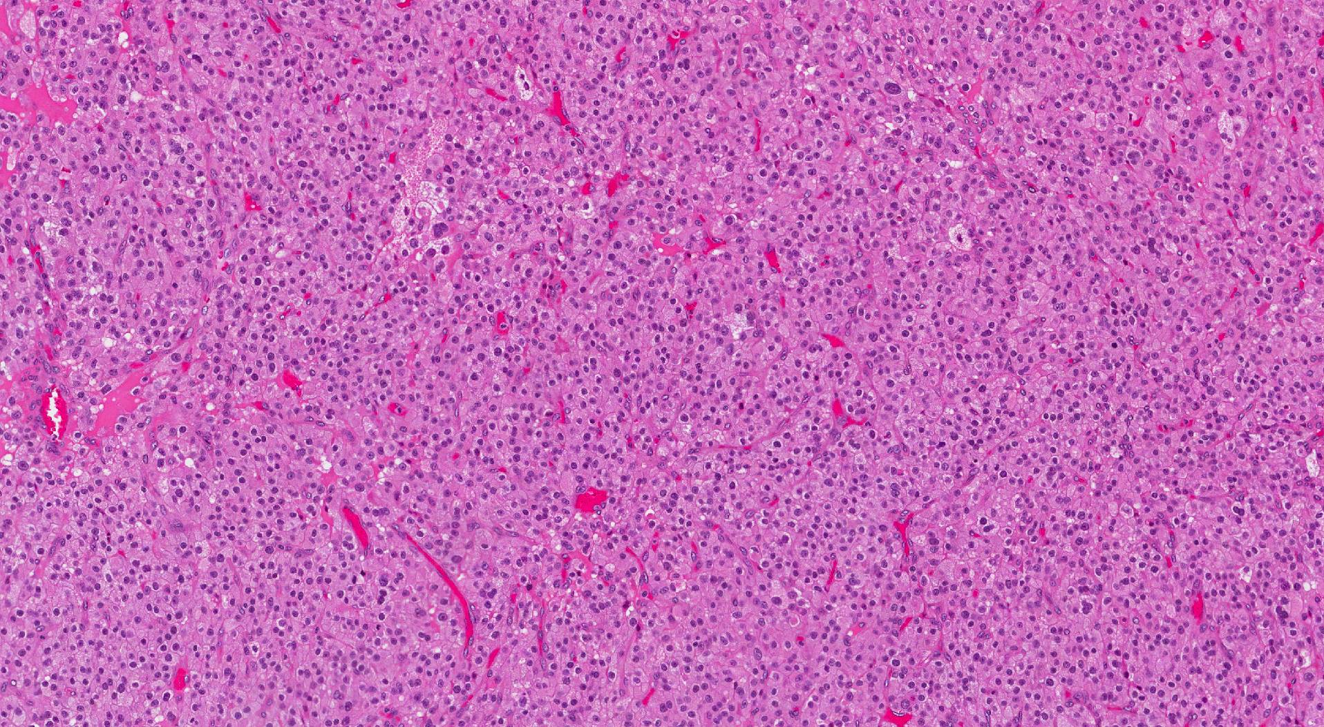
When examined under the microscope, most adrenal cortical adenomas are made up of cells that resemble the cells normally found in a part of the adrenal gland called the cortex. These cells are large and the cytoplasm (body) of the cell is full of lipid (fat) rich material. For this reason, the cells look white or clear when examined under the microscope. Some tumours also include smaller cells that contain less lipid and look eosinophilic (pink) when examined under the microscope. The tumour cells are usually arranged in small groups that are often described as cords, nests, or islands. Mitotic figures (tumour cells that are dividing to create new tumour cells) are very rarely seen and the tumour cells are usually surrounded by a thin layer of tissue called a tumour capsule.
Weiss score
The Weiss score is a system developed to help pathologists determine if an adrenal tumour is a non-cancerous adrenal cortical adenoma or a type of cancer called adrenocortical carcinoma. The Weiss score includes 9 features that pathologists look for when they are examining the tumour under the microscope. Each microscopic feature is given 1 point. A total score of less than 3 suggests that the tumour is unlikely to spread or behave like cancer and should be called an adrenal cortical adenoma. In contrast, a total score of 3 or more suggests that the tumour will behave like cancer and should be called adrenocortical carcinoma.
Here are the 9 features used to determine the Weiss score:
- High nuclear grade.
- More than mitotic figures (dividing tumour cells) 5 per 10mm2.
- Atypical mitotic figures.
- Necrosis (tumour cell death).
- Diffuse architecture (tumour cells growing in large groups).
- Clear cells make up less than 25% of the tumour.
- Tumour capsule invasion.
- Vascular invasion.
- Lymphatic invasion.

Do you have a question about your pathology report?
Our team of experts is here to help.
Ask a Pathologist.
Our work is generously supported by:
![]()