Basosquamous Carcinoma of the Skin: Understanding Your Pathology Report

Section Editor: Allison Osmond MD FRCPC
June 9, 2026


Print this article

Basosquamous carcinoma is a type of skin cancer. It is an uncommon, higher-risk form of basal cell carcinoma, the most common skin cancer. What makes basosquamous carcinoma different is that, in addition to the usual features of basal cell carcinoma, it also shows features of a second type of skin cancer called squamous cell carcinoma. Because of this mixed appearance, basosquamous carcinoma tends to behave somewhere between the two: it is more likely to invade deeper tissue, to recur after treatment, and, uncommonly, to spread compared with an ordinary basal cell carcinoma.

The tumor develops from basal cells, which are found in the lower part of the epidermis (the thin outer layer of the skin) and in hair follicles. This article explains what a diagnosis of basosquamous carcinoma means, what the findings in your pathology report describe, and how those findings guide the decisions you and your care team will make together. Most basosquamous carcinomas are cured when they are completely removed.

What causes basosquamous carcinoma?

The main cause of basosquamous carcinoma is long-term exposure to ultraviolet (UV) radiation, usually from the sun. Basal cells divide often to replace the surface of the skin, and this constant division makes them more likely to pick up the DNA damage that UV radiation causes. Over many years, this damage can build up and lead to cancer.

Other factors that raise the risk include fair skin that burns easily, older age, a weakened immune system (for example, after an organ transplant or with long-term immune-suppressing medication), and previous radiation therapy to the skin. People who have already had one skin cancer are at higher risk of developing another.

What are the symptoms of basosquamous carcinoma?

Basosquamous carcinoma usually appears as a slowly growing bump, firm patch, or sore on sun-exposed skin, most often on the head and neck. It can look like an ordinary basal cell carcinoma, so it often cannot be told apart from other skin cancers by eye alone. Common features include a raised or pearly bump, a scaly or crusted surface, an open sore that does not heal or that heals and reopens, and occasional bleeding after minor injury. Any new spot, or a spot that is changing, growing, or not healing, should be checked by a doctor.

How is the diagnosis made?

The diagnosis is made after a sample of the skin is examined under the microscope by a pathologist, a doctor who specializes in identifying disease in tissue. The sample is obtained by a skin biopsy. Depending on the size and location of the lesion, this may be a shave biopsy (a thin slice from the surface), a punch biopsy (a small round core of skin), or an excision (removal of the whole lesion).

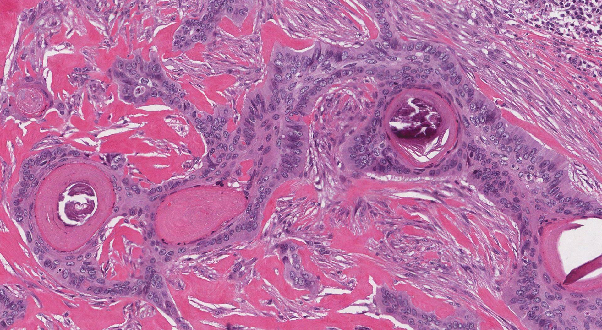
Under the microscope, basosquamous carcinoma shows two kinds of cells side by side. Part of the tumor looks like a typical basal cell carcinoma, with clusters of small, dark basaloid cells. Another part looks like squamous cell carcinoma, with larger cells that produce keratin, the tough protein found in the surface of normal skin. In many cases there is a transition zone where one pattern blends into the other. Because the tumor can resemble both an ordinary basal cell carcinoma and a pure squamous cell carcinoma, the pathologist may use immunohistochemistry (special stains that highlight particular proteins in the cells, such as a marker called Ber-EP4) to help confirm the diagnosis. If the diagnosis is made on a small biopsy, your doctor will usually recommend a second procedure to remove the rest of the tumor. Imaging is not needed for most small tumors, but it may be used for large, deep, or recurrent tumors to check how far the cancer has grown.

Histologic grade

Basosquamous carcinoma is not given a standard histologic grade (the well, moderately, or poorly differentiated scale used for many other cancers). Instead, the mixed basal cell and squamous cell appearance is itself what defines the tumor and marks it as a higher-risk form of basal cell carcinoma. For this reason, your pathology report will usually not include a grade number, and this is expected for this diagnosis.

Depth of invasion

Basosquamous carcinoma starts in the epidermis, the thin layer at the skin’s surface. Below the epidermis are the dermis (a thicker supporting layer) and the subcutaneous tissue (the fatty layer beneath the dermis). Depth of invasion describes how far the tumor cells have grown from the surface into the deeper layers, usually measured from the skin surface to the deepest tumor cells. Some reports describe this as tumor thickness. Tumors that grow more deeply are harder to remove completely and are more likely to come back or, uncommonly, to spread. The depth of invasion is usually reported only after the entire tumor has been removed.

Perineural invasion

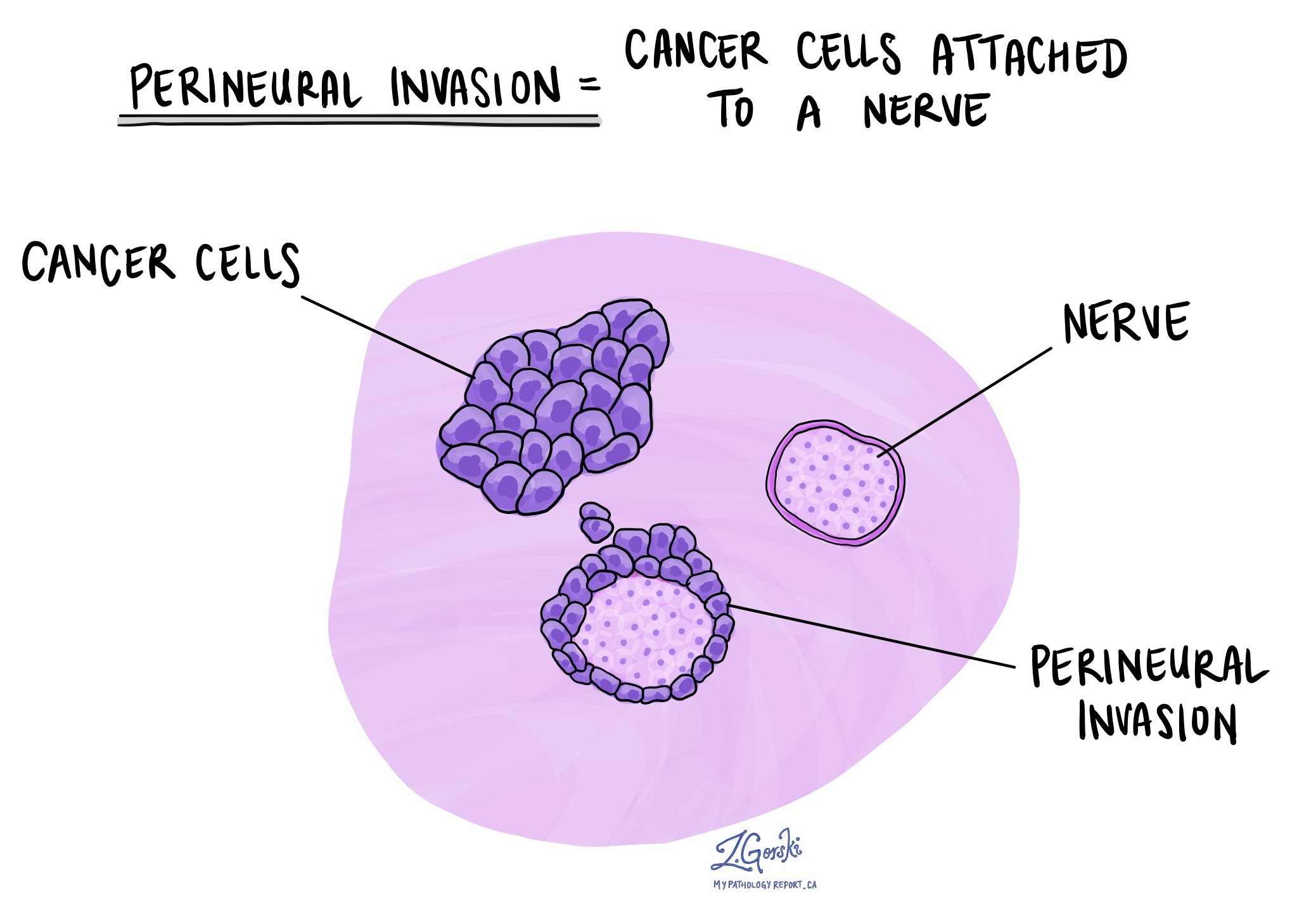
Perineural invasion means that tumor cells are seen attached to or growing along a nerve. Nerves run throughout the body and carry signals such as temperature, pressure, and pain. Perineural invasion is important because cancer cells can travel along a nerve into surrounding tissue, making the tumor harder to remove completely and increasing the risk that it will come back after treatment. This finding is more common in basosquamous carcinoma than in ordinary basal cell carcinoma, and when a large, named nerve is involved, it is considered a high-risk feature. Your report will state whether perineural invasion is present.

Lymphovascular invasion

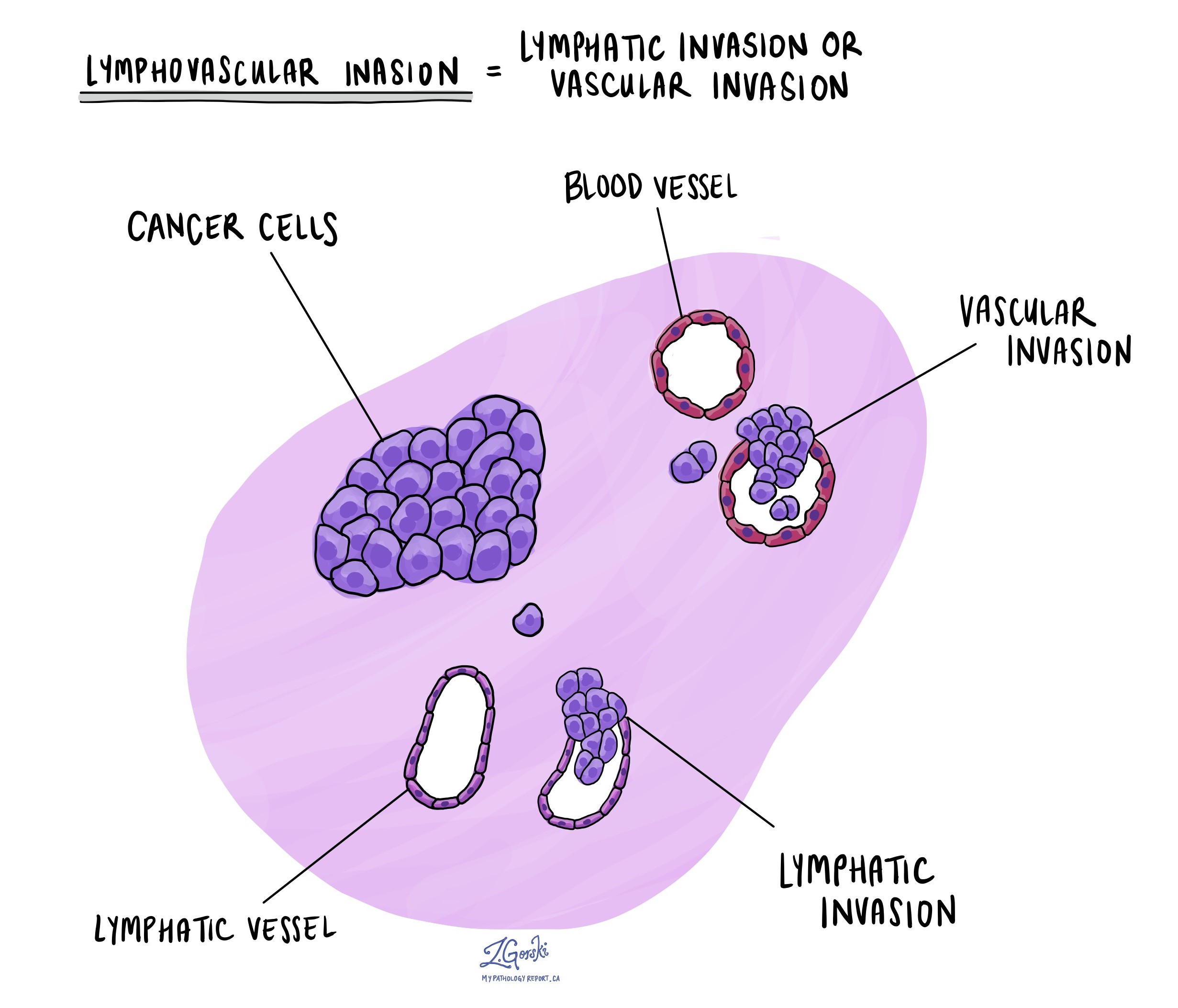
Lymphovascular invasion means that tumor cells are present within a blood or lymphatic vessel. Blood vessels carry blood around the body, while lymphatic vessels carry a clear fluid called lymph. These vessels are important because they provide cancer cells with a route to travel to lymph nodes or distant organs. Lymphovascular invasion is uncommon in basosquamous carcinoma, but when it is present, it raises the risk of spread, and your doctor may recommend closer follow-up or additional treatment. Your report will state whether lymphovascular invasion is present.

Surgical margins

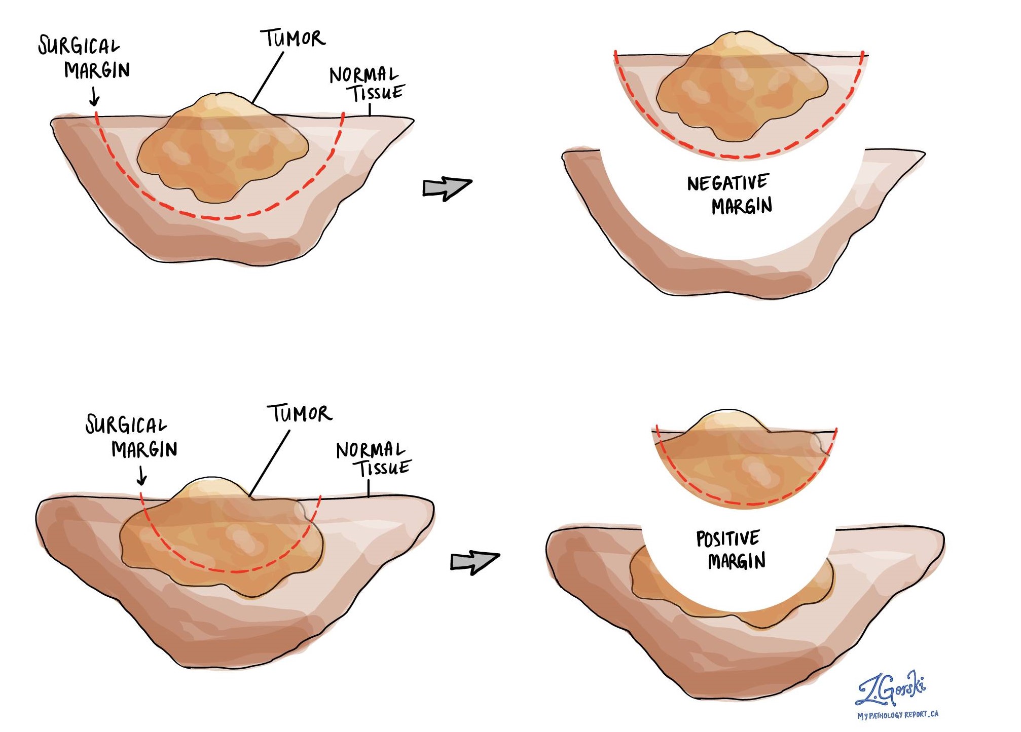
A margin is the rim of normal tissue removed around the tumor during surgery. Margins are usually reported only after an excision intended to remove the entire tumor, not after a small diagnostic biopsy. For basosquamous carcinoma, both the peripheral margin (the side edges of the removed skin) and the deep margin (the base of the removed tissue) are examined.

  • Negative margin — No tumor cells are seen at the cut edge. This suggests the tumor was completely removed. The pathologist may also record the distance from the nearest tumor cells to the edge, because a wider clear margin is associated with a lower risk that the tumor will return.
  • Close margin — Tumor cells are near the cut edge but do not reach it. Depending on how close they are, your doctor may discuss further surgery.
  • Positive margin — Tumor cells are present at the cut edge, which means some tumor may have been left behind. A positive margin is associated with a higher risk that the tumor will grow back at the same site, and further surgery or radiation may be considered.

Lymph nodes

Lymph nodes are small immune organs found throughout the body. Most basosquamous carcinomas never spread to lymph nodes, but the risk is higher than for an ordinary basal cell carcinoma. For this reason, lymph nodes are usually examined only when they feel enlarged or look suspicious on imaging. When that happens, a node may be sampled or removed and examined under the microscope. If a lymph node contains tumor, the report will state how many nodes were involved and the size of the largest deposit. For selected high-risk tumors, a sentinel lymph node biopsy (sampling of the first node that drains the area) may occasionally be considered.

Pathologic stage (pTNM)

Staging describes how large the tumor is and how far it has grown or spread. Most small basosquamous carcinomas that are completely removed are not assigned a formal stage. For larger or higher-risk tumors, particularly on the head and neck where most of these cancers occur, the AJCC (American Joint Committee on Cancer) staging system, 8th edition, for cutaneous carcinomas of the head and neck may be used. The tumor stage (pT) is based mainly on tumor size and whether the tumor has grown into deeper structures. Spread to distant organs (the M category) is determined by imaging rather than by the pathologist.

  • pT1 — Tumor is 2 cm or smaller.
  • pT2 — Tumor is larger than 2 cm but 4 cm or smaller.
  • pT3 — Tumor is larger than 4 cm, or there is minor erosion of nearby bone, perineural invasion of a named nerve, or deep invasion below the subcutaneous fat.
  • pT4 — Tumor has grown into deeper bone or the base of the skull.

What is the prognosis?

The outlook for basosquamous carcinoma is generally good, especially when the tumor is found early and completely removed. Most people are cured by surgery. Compared with an ordinary basal cell carcinoma, however, basosquamous carcinoma is more likely to come back after treatment and carries a higher (though still low) risk of spreading. Reported recurrence rates after standard surgical removal vary widely, from roughly 10% to 45%, and are substantially lower, around 4%, when the tumor is removed with Mohs micrographic surgery (a technique described below). Spread to other parts of the body is uncommon, occurring in about 5% of cases, and when it does happen it usually involves the nearby lymph nodes first.

Certain features in the pathology report are associated with a higher risk that the tumor will come back or spread:

  • Positive or close margins — Tumor cells at or near the cut edge increase the risk that the cancer will grow back at the same site.
  • Perineural invasion — Especially when a large, named nerve is involved.
  • Lymphovascular invasion — Indicates the tumor has reached vessels that can carry cells elsewhere.
  • Large tumor size and deep invasion — Larger and deeper tumors are harder to remove and more likely to recur.
  • High-risk location — Tumors on the central face, ears, lips, or scalp tend to behave less predictably.
  • Recurrent tumor — A cancer that has already returned once is more likely to return again.
  • Weakened immune system — People who are immunosuppressed are at higher risk of recurrence and spread.

What happens after the diagnosis?

The main treatment for basosquamous carcinoma is complete surgical removal. Because this tumor is more likely to recur than an ordinary basal cell carcinoma, the care team often considers Mohs micrographic surgery, a specialized technique in which the tumor is removed in thin layers that are checked under the microscope during the operation until no tumor cells remain at the edges. Mohs surgery offers the highest cure rate and is particularly useful for tumors on the head and neck and in areas where preserving healthy tissue matters. When Mohs surgery is not available or practical, a standard excision that removes the tumor with a rim of normal skin is used instead.

Radiation therapy may be considered after surgery when the report shows high-risk features, such as a positive margin that cannot be removed with more surgery or extensive perineural invasion. Radiation can also be the main treatment for people who are not candidates for surgery. In the uncommon situation in which basosquamous carcinoma has spread beyond the skin, the medical oncology team may consider systemic therapy. These can include hedgehog pathway inhibitors (drugs such as vismodegib and sonidegib used for advanced basal cell carcinoma) and immunotherapy drugs that help the immune system attack cancer; responses to these treatments vary, and the choice depends on the individual situation.

After treatment, regular skin examinations are important because people who have had basosquamous carcinoma are at higher risk of developing new skin cancers. Protecting the skin from the sun is a key part of long-term care. Care is often coordinated by a team that may include a dermatologist, a surgeon (often a Mohs surgeon), a radiation oncologist, a pathologist, and, for advanced disease, a medical oncologist.

Questions to ask your doctor

  • What type of skin cancer do I have, and how is basosquamous carcinoma different from an ordinary basal cell carcinoma?
  • How large was the tumor, and how deep had it grown into the skin?
  • Was the whole tumor removed, or were tumor cells found at the margins?
  • If the margins were positive, do I need more surgery or radiation?
  • Was perineural invasion or lymphovascular invasion seen in my tumor?
  • Would Mohs micrographic surgery be a good option for removing this tumor?
  • Is my tumor in a high-risk location, such as the face, ears, or lips?
  • What is the chance that this cancer will come back at the same place?
  • What is the chance it has spread to lymph nodes or elsewhere?
  • Do I need any imaging or lymph node evaluation?
  • How often should I have my skin checked, and what changes should I watch for?
  • Am I at higher risk because of a weakened immune system or previous radiation?

Related articles

A+ A A-
Was this article helpful?