

by Jason Wasserman MD PhD FRCPC
August 10, 2023
What is a fibrothecoma?
A fibrothecoma is an uncommon and noncancerous type of ovarian tumour made up of thecal cells and fibroblastic spindle cells. It shares features with two other types of noncancerous ovarian tumours, thecomas and fibromas.
Is a fibrothecoma benign or malignant?
Fibrothecoma is a benign (noncancerous) type of ovarian tumour.
What is the difference between a fibrothecoma and a fibroma?
Fibrothecoma and fibroma are very similar tumours. The main difference is that a fibrothecoma is made up of both thecal cells and spindle cells while a fibroma is made up only of spindle cells. Both tumours are benign (noncancerous).
What are the symptoms of a fibrothecoma?
The most common symptoms of fibrothecoma are related to excess hormone production by the tumour. Most tumours produce excess estrogenic hormones, however, symptoms related to excess androgen production are also possible. Large tumours may cause symptoms such as pelvic or abdominal pain and pressure.
What causes a fibrothecoma?
At present, we do not know what causes a fibrothecoma.
How is this diagnosis made?
The diagnosis is made after the entire tumour has been surgically removed and examined under the microscope by a pathologist.
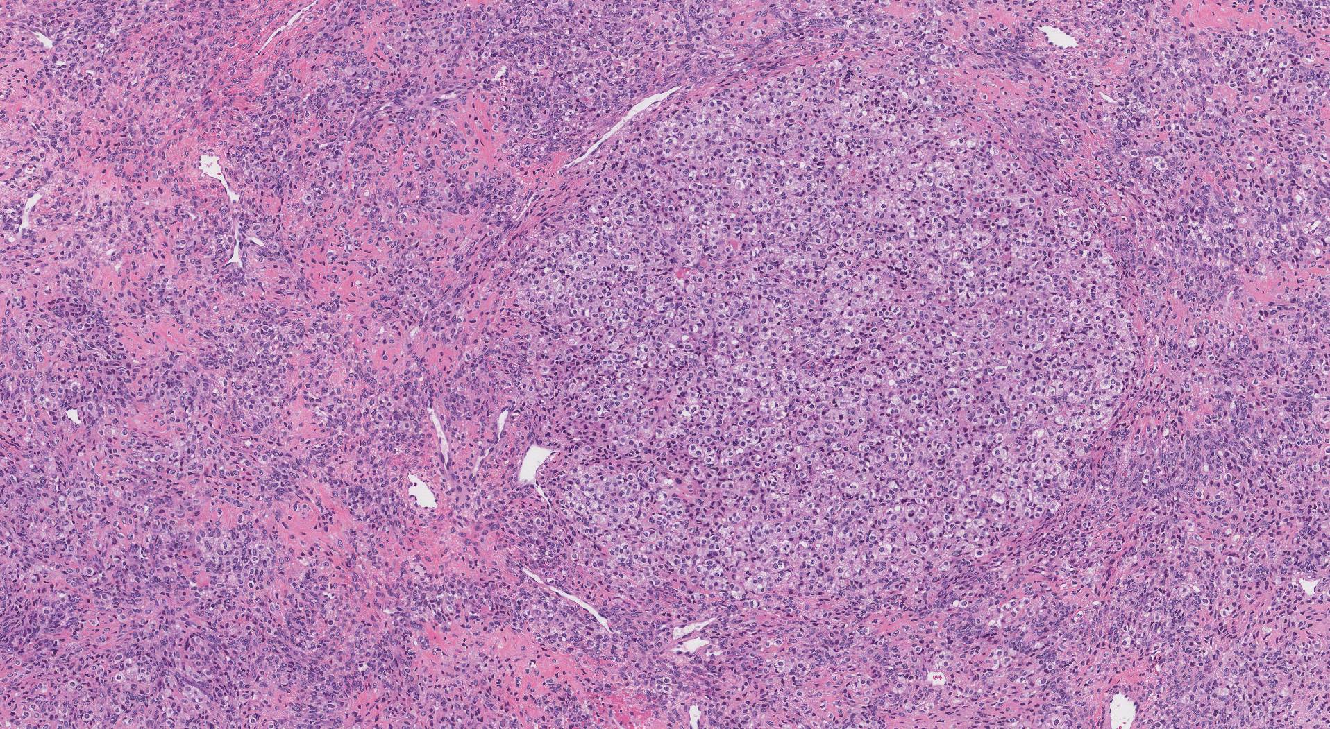
What does a fibrothecoma look like under the microscope?
When examined under the microscope, fibrothecomas are made up of two types of cells: thecal cells and fibroblastic spindle cells. The thecal cells are large cells with pink-grey coloured cytoplasm and round nuclei. These cells are typically arranged in nests or large groups called sheets. A tumour made up entirely of thecal cells is called a thecoma. The fibroblastic spindle cells in a fibrothecoma are long, thin cells that look very similar to the cells found in an ovarian fibroma. The spindle cells tend to be surrounded by a collagenous or fibrotic stroma. Mitotic figures (cells dividing to create new cells) may be seen but should be rare.
We are proud to partner with:
![]()