

by Glenda Wright MBBCh and Allison Osmond MD FRCPC
July 25, 2024
Background:
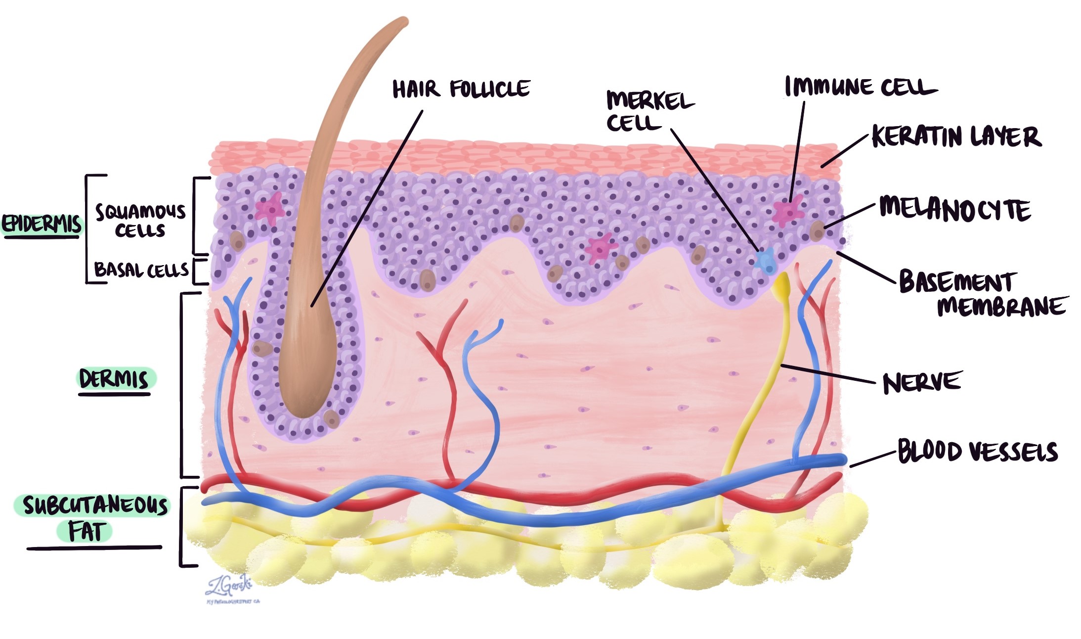
Fibrous histiocytoma is a common benign (non-cancerous) skin tumour. It is made up of a combination of fibroblastic cells, macrophages, and collagen, a type of connective tissue. The tumour develops in a layer of skin called the dermis. Another name for a fibrous histiocytoma is dermatofibroma.
What are the symptoms of a fibrous histiocytoma?
Most fibrous histiocytomas grow slowly, have a pink to brown colour, and are usually small and painless.
What causes a fibrous histiocytoma?
Some fibrous histiocytomas appear to develop after trauma, an insect bite, or prolonged inflammation of the skin. However, most tumours develop without any preceding event and the cause remains unknown.
Can a fibrous histiocytoma turn into cancer over time?
No. A fibrous histiocytoma is a benign (non-cancerous) tumour that will not become cancerous over time. However, tumours that are not completely removed can re-grow.
Subtypes of fibrous histiocytoma
Several subtypes of fibrous histiocytoma exist, each with distinct microscopic and clinical features.
Aneurysmal fibrous histiocytoma
Aneurysmal fibrous histiocytoma is characterized by large, blood-filled spaces within the tumor, resembling an aneurysm. These spaces are lined by fibrohistiocytic cells and often contain hemosiderin-laden macrophages. The surrounding stroma shows spindle cells and a mix of inflammatory cells.
The aneurysmal fibrous subtype typically presents as a rapidly growing, red-to-bluish nodule, often on the extremities. Due to its hemorrhagic appearance, it may resemble a vascular lesion.
Cellular fibrous histiocytoma
Cellular fibrous histiocytoma shows increased cellularity with dense, spindle-shaped fibroblasts and histiocytes arranged in a storiform pattern. Mitotic figures may be more frequent, and there is often a peripheral collagenous rim.
Cellular fibrous histiocytomas are usually firm, flesh-colored nodules that can appear on any part of the body but are more common on the legs. They may be larger and more painful compared to typical fibrous histiocytomas.
Deep fibrous histiocytoma
Deep fibrous histiocytoma extends into the subcutaneous tissue or deeper, involving the fascia or muscle. It retains the typical storiform pattern of fibroblasts and histiocytes but with a deeper extension.
Deep fibrous histiocytomas are often larger and may present as deep-seated, firm nodules. Due to their size and depth, they can be mistaken for malignant soft tissue tumors.
Clear cell fibrous histiocytoma
Clear cell fibrous histiocytoma contains clear cells with abundant, pale-staining cytoplasm, which are intermixed with typical fibrohistiocytic cells. The clear cells are often glycogen-rich and PAS-positive.
This rare subtype typically presents as a slow-growing, painless nodule, often on the extremities. Its clear cell features can resemble other clear cell neoplasms, necessitating careful histological evaluation.
Lipidized fibrous histiocytoma
Lipidized fibrous histiocytoma is characterized by the presence of lipid-containing foam cells (lipidized histiocytes) within the tumour. These foam cells are mixed with spindle cells and other histiocytes.
Lipidized fibrous histiocytomas are usually yellowish or tan nodules, reflecting the presence of lipid-laden cells. They are most commonly found on the lower extremities.
Palisaded fibrous histiocytoma
Palisaded fibrous histiocytoma features spindle cells arranged in a palisading pattern around central areas of collagen or necrosis. The tumour also includes a mix of histiocytes and inflammatory cells.
This subtype typically presents as a firm, skin-colored, or slightly erythematous nodule. It is most commonly found on the extremities or trunk.
Myxoid fibrous histiocytoma
Myxoid fibrous histiocytoma contains abundant myxoid stroma, which is a gelatinous matrix. Spindle cells and histiocytes are dispersed within this myxoid background, often with a storiform or loose fascicular pattern.
Due to the myxoid matrix, myxoid fibrous histiocytomas usually present as soft, sometimes cystic nodules on the extremities and may be mistaken for myxoid soft tissue tumors.
Epithelioid fibrous histiocytoma
Epithelioid fibrous histiocytoma is characterized by large, polygonal cells with abundant eosinophilic cytoplasm that resemble epithelial cells. These epithelioid cells are arranged in nests or sheets and are often surrounded by typical spindle-shaped fibroblasts and histiocytes.
Epithelioid fibrous histiocytomas typically present as small, firm, red to brown nodules on the skin, most commonly on the extremities. They may resemble other epithelioid lesions, such as Spitz nevi or melanomas, making histological evaluation important for accurate diagnosis.
How is this diagnosis made?
The diagnosis is usually made after the entire tumour has been surgically removed and sent to a pathologist for examination under a microscope. Sometimes, your doctor will remove a small piece of the tumour in a procedure called a biopsy before removing the entire tumour.
About this article
Doctors wrote this article to help you read and understand your pathology report. If you have additional questions, contact us.
Other helpful resources
Atlas of Pathology
We are proud to partner with:
![]()