

by Bibianna Purgina, MD FRCPC
October 6, 2022
What is a pleomorphic lipoma?
A pleomorphic lipoma is a non-cancerous tumour made up of fat and spindle cells. This tumour commonly starts around the upper back and neck but can be found almost anywhere in the body. Most grow just under our skin where they feel like a soft lump. Another name for this tumour is spindle cell lipoma.
What are the symptoms of pleomorphic lipoma?
Most pleomorphic lipomas are slow-growing and cause very few symptoms. Large tumours on the back may cause pain caused by pressure.
What causes pleomorphic lipoma?
At present doctors do not know what causes pleomorphic lipoma.
How is the diagnosis of pleomorphic lipoma made?
The diagnosis is made after part or all of the tumour is removed and sent to a pathologist for examination under the microscope.
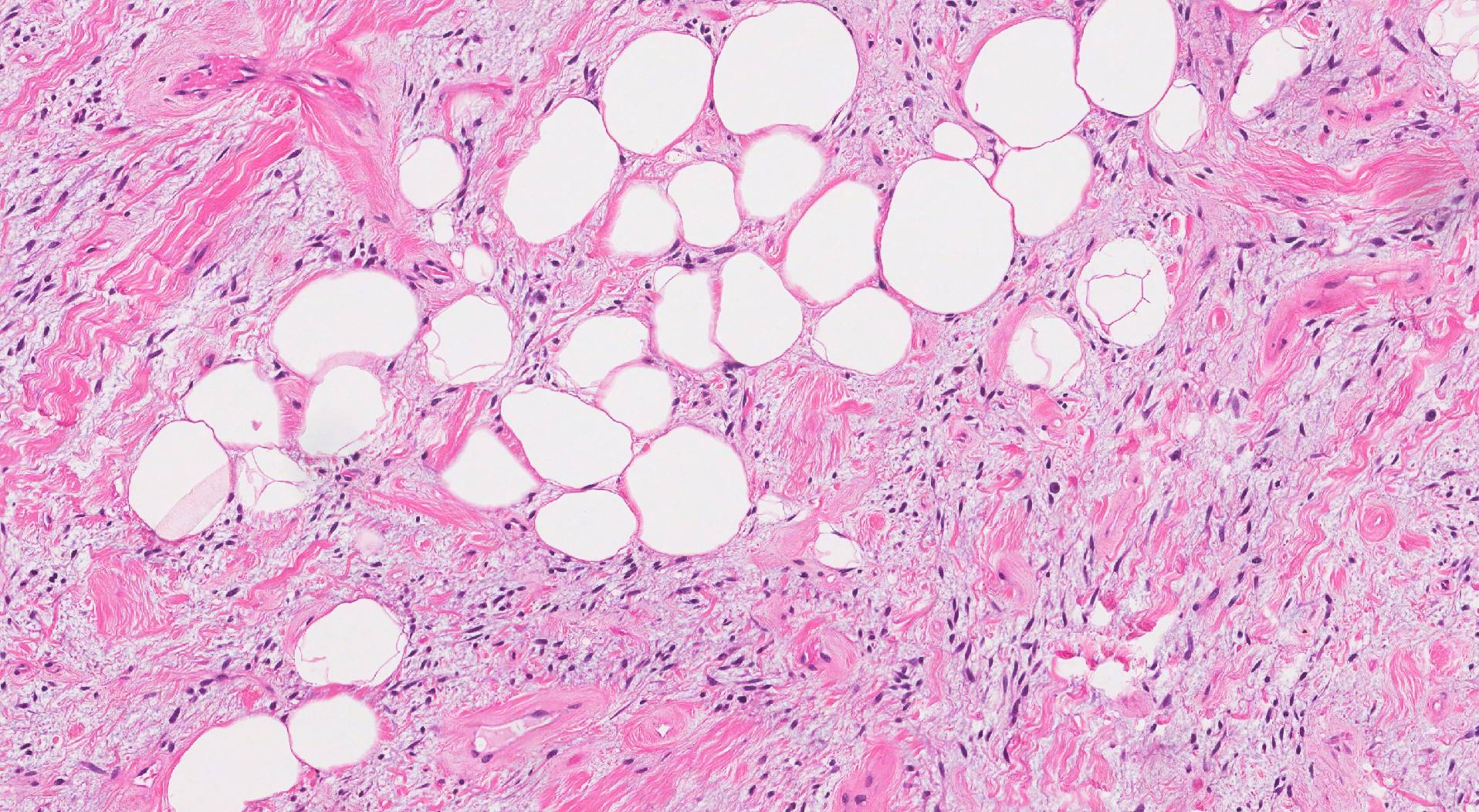
What does a pleomorphic lipoma look like under the microscope?
When examined under the microscope, the tumour is made up of a combination of adipocytes (fat cells) and spindle cells. The adipocytes in a pleomorphic lipoma look almost identical to normal adipocytes found in fat throughout the body. However, in a pleomorphic lipoma, the adipocytes are surrounded by long, thin spindle cells. Pathologists describe these spindle cells as ‘pleomorphic’ because they range in shape and size. Multinucleated tumour cells (tumour cells with more than one nucleus) may also be seen with some tumours being predominantly multinucleated cells. The stroma or connective tissue holding the tumour together often looks very pink under the microscope.
What other tests may be performed to confirm the diagnosis?
Your pathologists may perform additional tests including immunohistochemistry and fluorescence in situ hybridization (FISH) to confirm the diagnosis and to exclude other tumours that can look like a pleomorphic lipoma.
Immunohistochemistry
Immunohistochemistry is a test that allows pathologists to see specific types of chemicals such as proteins inside cells. When this test is performed, the tumour cells in a pleomorphic lipoma are usually positive (reactive) for S100 and CD34.
Fluorescence in situ hybridization (FISH) for MDM2
MDM2 is a gene that promotes cell division (the creation of new cells). Normal cells and those in pleomorphic lipomas have two copies of the MDM2 gene. In contrast, some cancers that look like pleomorphic lipomas have more than two copies of the MDM2 gene.
Fluorescence in situ hybridization (FISH) is a test that allows pathologists to count the number of gene copies in a cell. A normal copy number or a normal ratio of MDM2 to a control gene such as CEP12 confirms that the tumour is a non-cancerous pleomorphic lipoma.
We are proud to partner with:
![]()