

by Rosemarie Tremblay-LeMay MD FRCPC
May 1, 2024
Thrombotic microangiopathy (TMA) is a group of conditions where red blood cells (RBCs) are destroyed by blood clots in small blood vessels such as capillaries and arterioles. TMA is also associated with decreased platelets and organ damage.
Diseases in this group include:
- Disseminated intravascular coagulation (DIC)
- Thrombotic thrombocytopenic purpura (TTP)
- Hemolytic uremic syndrome (HUS)
- TMA occurring after bone marrow transplantation
What causes thrombotic microangiopathy?
TMA is caused by the uncontrolled formation of small blood clots in small blood vessels, called thrombi. The blood clots block the inside of the blood vessel and damage or destroy the RBCs as they try to pass. Pathologists describe this process as non-immune hemolysis because the immune system is not involved in damaging the RBCs. Conditions that can lead to the uncontrolled formation of blood clots and TMA include infections, cancer, certain types of medications, pregnancy, organ transplantation, or trauma.

How do doctors test for thrombotic microangiopathy?
Your doctor will perform blood tests to look for substances released from damaged RBCs, such as indirect bilirubin and LDH. There will also be a decrease in haptoglobin. Depending on the type of TMA, tests that measure substances involved in blot clot formation may also be abnormal. Some tests can help determine if other organs are affected, such as the kidneys.
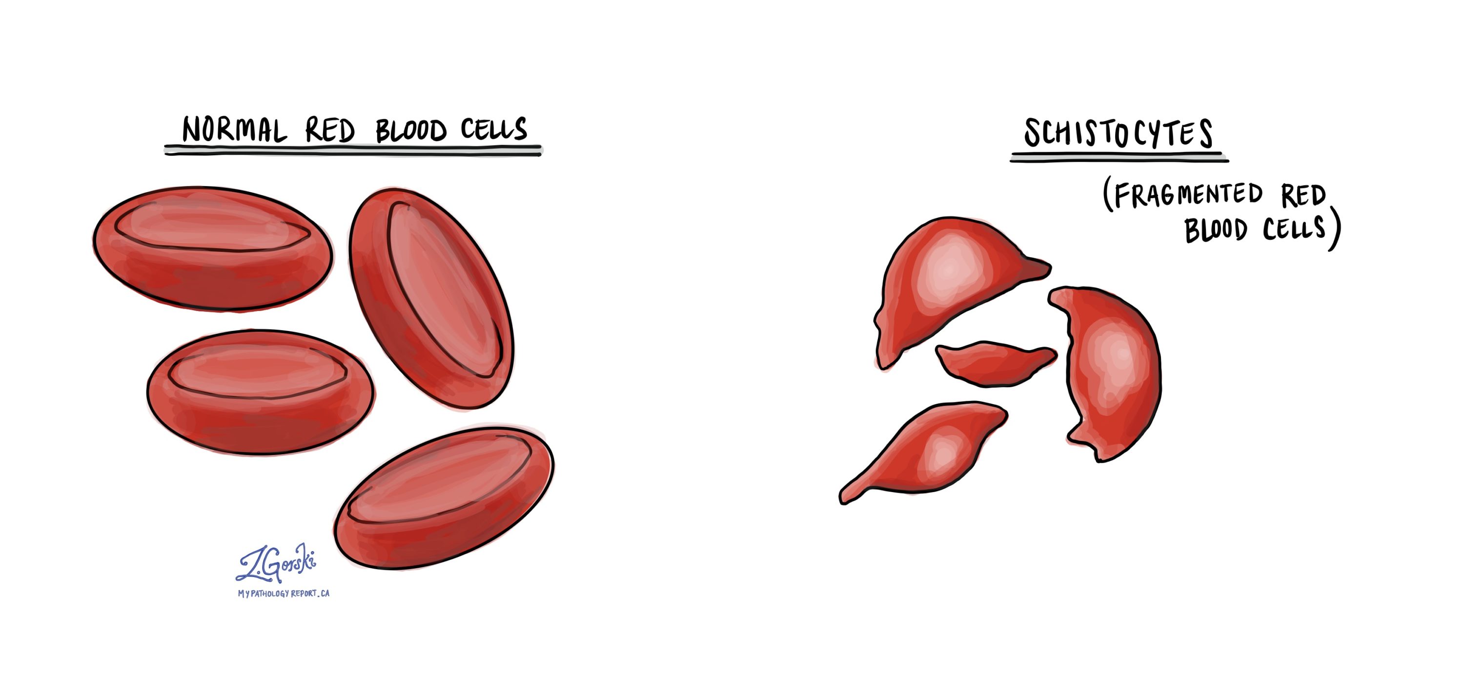
What does thrombotic microangiopathy look like under the microscope?
When a blood sample is examined under the microscope, small fragments of damaged RBCs, or schistocytes, can be seen. There will also be an increased number of immature RBCs and a decreased number of platelets.
About this article
Doctors wrote this article to help you read and understand your pathology report. Contact us if you have any questions about this article or your pathology report. Read this article for a more general introduction to the parts of a typical pathology report.
Other helpful resources
Atlas of Pathology
We are proud to partner with:
![]()