

Jason Wasserman MD PhD FRCPC
August 10, 2024
A xanthoma is a type of growth that forms under the skin or in other tissues of the body. It is made up of fat-filled cells called foamy histiocytes, which are a type of immune cell that has absorbed fat. Xanthomas can appear anywhere on the body but are most commonly found on the skin. They are generally not harmful but may indicate an underlying medical condition, such as high cholesterol levels.
What are the symptoms of a xanthoma?
The symptoms of a xanthoma can vary depending on its location and size. Xanthomas often appear as yellowish or orange bumps or patches on the skin. They are usually not painful, but they can be itchy or tender to the touch. In some cases, xanthomas may be found in deeper tissues, such as tendons, where they may not cause any noticeable symptoms.
What causes a xanthoma?
Xanthomas are caused by fat accumulation in certain cells within the body. This can happen for several reasons, including high levels of cholesterol or other fats (lipids) in the blood. Conditions that lead to high lipid levels, such as familial hypercholesterolemia (a genetic condition that causes high cholesterol), diabetes, and certain liver diseases, can increase the risk of developing xanthomas. In some cases, xanthomas may also occur without any underlying health problems.
Types of xanthomas
There are several types of xanthomas, each with its own unique features:
- Xanthelasma: This is the most common type of xanthoma and appears as yellowish patches on the eyelids. It is usually seen in older adults and is often associated with high cholesterol levels.
- Palmar xanthoma: These xanthomas occur on the palms of the hands. They are typically flat and may be associated with underlying lipid disorders.
- Tendon xanthoma: These xanthomas form in the tendons, which are the tissues that connect muscles to bones. They are most commonly found in the Achilles tendon and the tendons of the hands. Tendon xanthomas are often associated with high cholesterol levels.
- Tuberous xanthoma: These are firm, painless nodules that typically appear on the knees, elbows, or buttocks. They are often associated with high levels of cholesterol or triglycerides in the blood.
- Verruciform xanthoma: This rare type of xanthoma usually appears as a wart-like lesion on the skin, most commonly in the mouth or genital area. It is not typically associated with lipid disorders.
- Eruptive xanthoma: These xanthomas appear as small, red-yellow bumps that suddenly appear on the skin, often on the buttocks, shoulders, or thighs. They are usually associated with very high blood triglyceride levels.
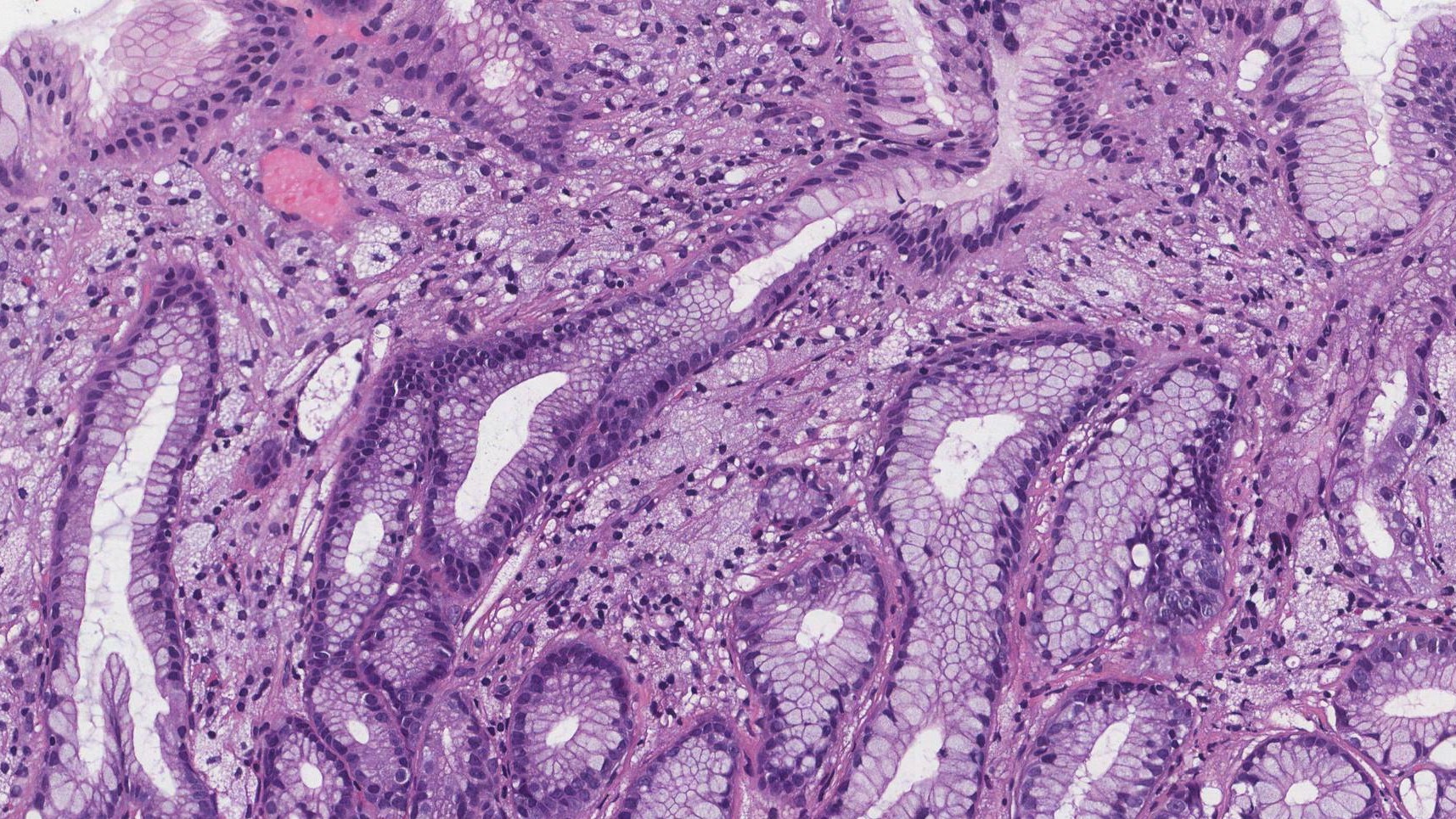
What does a xanthoma look like under the microscope?
Under the microscope, a xanthoma is made up of a collection of foamy histiocytes. These large cells are filled with fat, which gives them a foamy appearance. The foam cells are often surrounded by other types of immune cells. In some types of xanthomas, such as tendon xanthomas, the growth may also contain areas of calcification (deposits of calcium) or fibrosis (thickening and scarring of connective tissue).
We are proud to partner with:
![]()