

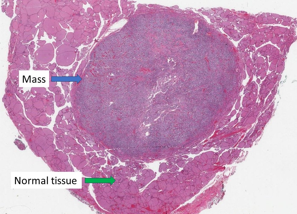
The word “mass” in a pathology report refers to an area of tissue that is larger than normal. It is a broad term used to describe a lump or abnormal growth that can be seen or felt during a physical examination or detected using imaging tests like an X-ray or MRI. A mass can occur anywhere in the body and may vary in size and appearance.
In medical terms, a mass does not automatically mean cancer. It is simply a descriptive term for something that appears larger than it should in a particular body part.
What types of conditions can cause a mass?
Many different conditions can cause a mass, and not all of them are harmful. Common causes include:
- Infections: An infection can lead to swelling or the formation of an abscess, which is a pocket of pus.
- Inflammation: Inflammatory conditions, like arthritis or autoimmune diseases, can cause localized swelling or enlargement of tissue.
- Cysts: A cyst is a fluid-filled sac that can develop in various parts of the body, such as the skin, ovaries, or liver.
- Benign tumours: These are non-cancerous growths that can form in tissues like muscle, fat, or bone.
- Malignant tumours: These are cancerous growths that can invade nearby tissues and spread to other parts of the body.
Sometimes, a mass is caused by normal body changes, such as a swollen lymph node during a cold or scar tissue forming after an injury.
Is a mass always cancer?
No, a mass is not always cancer. In fact, many are non-cancerous (benign) and do not pose a serious health risk. Examples include lipomas (fatty growths), fibroids (common in the uterus), and simple cysts.
Cancerous (malignant) masses, on the other hand, have the potential to grow and spread to other parts of the body. Because of this, it is important to investigate any new or unusual mass to determine its cause. However, the majority of masses found in routine examinations turn out to be non-cancerous.
How is this diagnosis made?
Diagnosing the cause of a mass typically involves several steps:
- Physical examination: Your doctor may examine the mass by feeling it to assess its size, texture, and whether it is fixed or movable.
- Imaging tests: Tests like X-rays, CT scans, or MRIs provide detailed pictures of the mass and surrounding tissues.
- Laboratory tests: Blood tests or other lab work may help identify markers that point to specific conditions.
- Biopsy: If more information is needed, a small piece of the mass may be removed and examined under a microscope by a pathologist. This is called a biopsy and is the most reliable way to determine whether a mass is benign or malignant.
By combining these methods, doctors can determine the nature of the mass and recommend the appropriate next steps.
About this article
This article was written by doctors to help you read and understand your pathology report. Contact us if you have questions about this article or your pathology report. For a complete introduction to your pathology report, read this article.
We are proud to partner with:
![]()