

In pathology, “grade” is a term used to describe the appearance and behavior of cells within a tissue sample, typically in the context of cancerous tumors and precancerous conditions, as well as some noncancerous conditions. Grade is important because it provides clues about the potential for disease progression and guides treatment decisions.
Grade of cancer
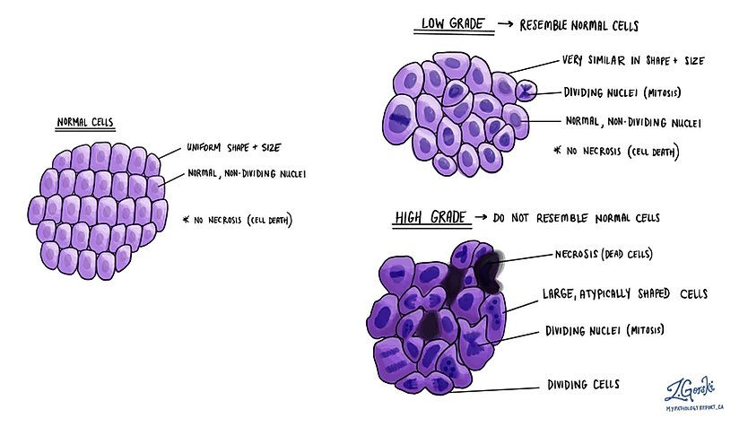
In the context of cancer, grade refers to the evaluation of how much tumor cells resemble the normal cells from which they originated. This assessment, performed under a microscope by a pathologist, considers the level of differentiation of the cancer cells—the extent to which these abnormal cells retain the characteristics of their original tissue.
- Low grade: Cells that look similar to normal cells and tend to grow slowly. This grade is sometimes described as well differentiated.
- Intermediate grade: Cells that exhibit more abnormalities and have a growth rate and potential to spread that is intermediate between low and high grades. This grade is sometimes described as moderately differentiated.
- High grade: Highly abnormal cells that grow and spread quickly, showing little resemblance to the original tissue. This grade is sometimes described as poorly differentiated.
The grade of a cancer plays a vital role in determining the aggressiveness of the disease and influences both prognosis and treatment strategies.
Grade in precancerous conditions: dysplasia
Dysplasia is the abnormal development or growth of cells within tissues and is often considered a precursor to cancer. Pathologists grade dysplasia by assessing the extent of cellular abnormalities and architectural disruption, which helps predict the risk of progression to cancer.
- Mild dysplasia: Cells are slightly abnormal and are at a lower risk of progressing to cancer. Mild dysplasia is sometimes described as low grade dysplasia.
- Moderate dysplasia: Cells show a higher degree of abnormality, with an increased risk of developing into cancer compared to mild dysplasia. Depending on the area of the body involved, moderate dysplasia can be described as either low grade or high grade dysplasia.
- Severe dysplasia: Cells are significantly abnormal and closely resemble cancerous cells, representing a high risk of progression to invasive cancer. Severe dysplasia is sometimes described as high grade dysplasia.
The grading of dysplasia is particularly important in conditions like cervical intraepithelial neoplasia (CIN), where it guides the monitoring and treatment approach to prevent the development of cervical cancer.
Grade in noncancerous conditions
In some noncancerous conditions, grading can be used to describe the severity or extent of tissue abnormality. For example, grade can be applied to inflammatory conditions, where it may reflect the degree of tissue damage or inflammation. Although not universally used in the same way as in cancer or precancerous conditions, understanding the grade in these contexts can help in assessing disease severity and planning treatment.
We are proud to partner with:
![]()