

Reviewed by Pathologists on:
January 8, 2026
Myoepithelial cells are specialized cells that have features of both muscle cells and epithelial cells. Their primary role is to help push secretions out of glands. They do this by gently contracting, similar to muscle cells, while also forming part of the gland structure, like epithelial cells.
These cells are essential to many normal glands and play a key role in how those glands function.
Where are myoepithelial cells normally found?
Myoepithelial cells are found in several types of glands throughout the body.
They are most commonly seen in:
-
Salivary glands, where they help release saliva.
-
Breast tissue, where they help move milk through the ducts.
-
Sweat glands, where they assist in pushing sweat to the skin surface.
-
Lacrimal glands, which produce tears.
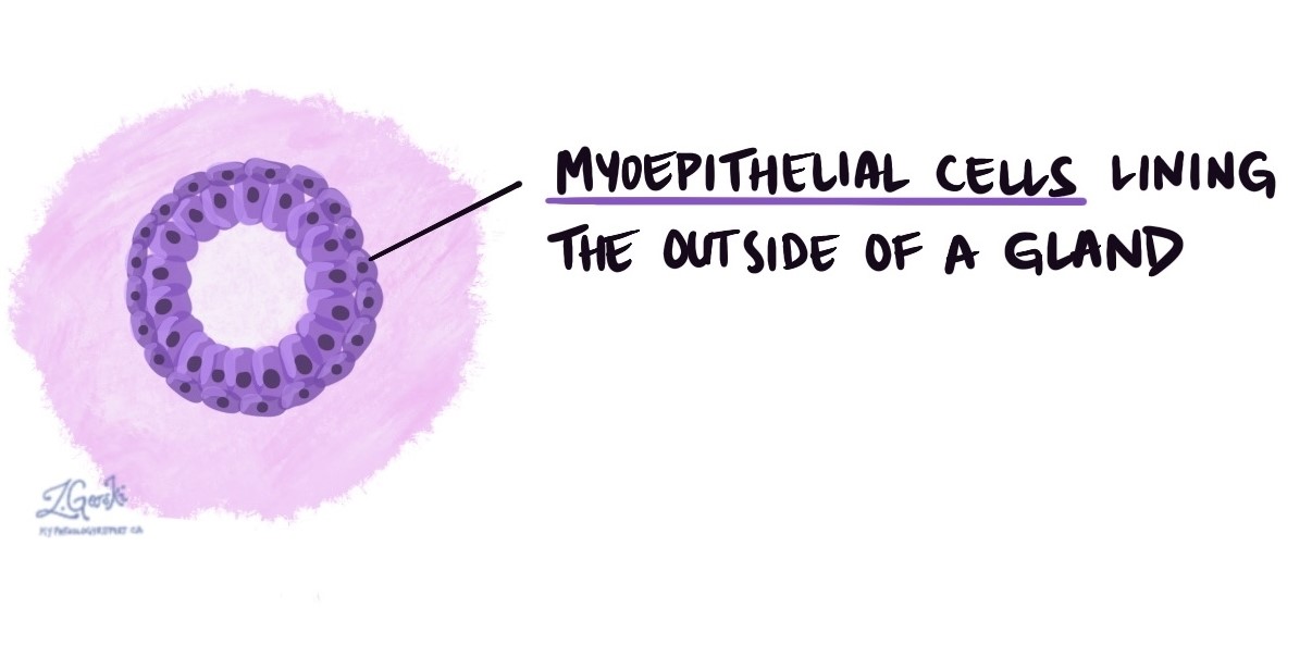
In these locations, myoepithelial cells sit between the glandular cells and the surrounding connective tissue, forming a thin supportive layer around ducts and glandular units.
What do myoepithelial cells do?
The primary function of myoepithelial cells is to contract and squeeze glandular structures, helping secretions move from the gland into ducts and toward the surface. This action supports normal gland function and helps keep ducts open.
Myoepithelial cells also help maintain the normal structure of glands and act as a partial barrier between gland cells and surrounding tissue.
What do myoepithelial cells look like under the microscope?
Under the microscope, myoepithelial cells are usually flattened or spindle-shaped and form a thin layer around ducts or glandular units. They may be difficult to see on routine stains because they are small and blend in with the surrounding tissue.
Pathologists often use immunohistochemical staining to highlight myoepithelial cells. These stains detect proteins that myoepithelial cells normally produce, making them easier to identify.
Why are myoepithelial cells important in pathology?
Myoepithelial cells are essential in pathology because their presence or absence can help pathologists determine whether a glandular lesion is benign (non-cancerous) or malignant (cancerous).
In many benign glandular conditions, myoepithelial cells are preserved. In contrast, invasive cancers often lose the myoepithelial cell layer, allowing tumour cells to spread into surrounding tissue. For this reason, identifying myoepithelial cells can help pathologists assess whether a tumour is confined to its normal location or has begun to invade.
What conditions are associated with myoepithelial cells?
Myoepithelial cells may be mentioned in pathology reports describing:
-
Benign tumours, such as pleomorphic adenoma of the salivary gland.
-
Myoepithelial tumours, which are made up mostly or entirely of myoepithelial cells.
-
Breast lesions, where the presence or absence of myoepithelial cells helps distinguish in situ (non-invasive) from invasive cancer.
The interpretation depends on the tissue type and the overall microscopic findings.
What cancers show a loss of myoepithelial cells?
Loss of myoepithelial cells is a key feature of invasive glandular cancers. When these cells are no longer present around ducts or glands, it suggests that cancer cells have broken through their normal boundaries.
Examples include:
- Invasive breast carcinoma, where the loss of myoepithelial cells helps distinguish invasive cancer from non-invasive conditions such as ductal carcinoma in situ (DCIS).
- Invasive salivary gland carcinomas, such as salivary duct carcinoma or adenocarcinoma, which typically lack a surrounding myoepithelial cell layer.
- Invasive sweat gland carcinomas, where the disappearance of myoepithelial cells supports a diagnosis of malignancy.
In contrast, some cancers called myoepithelial carcinomas are composed of malignant myoepithelial cells. In these cases, myoepithelial markers are present, but the normal organized myoepithelial layer is lost.
Identifying whether myoepithelial cells are present, absent, or abnormally arranged helps pathologists determine whether a tumour is invasive and guides diagnosis and treatment.
Why might myoepithelial cells be mentioned in my pathology report?
Your pathology report may mention myoepithelial cells to explain how a diagnosis was made. The report may note whether these cells are present, reduced, or absent around glands or ducts.
This information helps your doctor understand whether the tissue shows normal gland structure, a benign condition, or features concerning for cancer.
Questions to ask your doctor
-
Were myoepithelial cells seen in my tissue sample?
-
Does their presence or absence affect my diagnosis?
-
Were special stains used to identify these cells?
-
What does this finding mean for my treatment or follow-up?
We are proud to partner with:
![]()