

In pathology, the term nonkeratinizing describes squamous cells that have not undergone keratinization. Keratinization is a natural process where cells produce and store a tough, protective protein called keratin. When squamous cells do not go through this process, they are called nonkeratinizing.
Nonkeratinizing squamous cells are normally found in moist mucosal tissues throughout the body. These cells help protect the underlying tissue, reduce friction, and keep the surface soft and flexible. Common sites with nonkeratinizing squamous cells include the cervix, oral cavity, oropharynx, esophagus, anus, and nasal cavity.
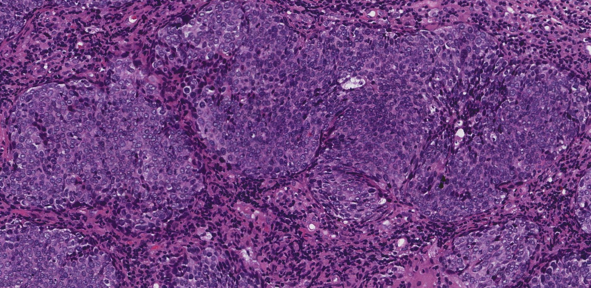
What do nonkeratinizing cells look like under the microscope?
Pathologists use a special stain called hematoxylin and eosin (H&E) to look at cells under the microscope. When stained this way, nonkeratinizing squamous cells usually appear dark blue or purple. This is because they have a large nucleus, the part of the cell that contains the genetic material, and the nucleus takes up most of the space inside the cell. These cells do not have the pink, dense appearance seen in keratinizing squamous cells, which are packed with keratin in the cytoplasm (the body of the cell).
In contrast, keratinizing squamous cells look bright pink due to the buildup of keratin in the cytoplasm. These cells often have a small, shrunken nucleus or no nucleus at all if they are fully keratinized.
Where are nonkeratinizing cells normally found in the body?
Nonkeratinizing squamous cells are part of the normal lining of many mucosal surfaces, which are areas that remain moist and exposed to air or fluids. These include:
-
The cervix (especially the transformation zone).
-
The vagina.
- The oral cavity including the mouth and tongue.
-
The oropharynx (tonsils, soft palate, base of tongue).
-
The nasal cavity and nasopharynx.
-
The esophagus.
-
The anus.
-
Parts of the urinary tract.
These cells help form a protective barrier while staying soft and flexible, unlike the tough outer layers of the skin, which are made of keratinizing squamous cells.
What does it mean if a tumour is described as nonkeratinizing?
When a tumour is described as nonkeratinizing, it means the tumour cells resemble nonkeratinizing squamous cells. These cells do not produce keratin, and they usually maintain a large, visible nucleus with less pink cytoplasm. This information helps pathologists determine what kind of cancer is present.
The most common type of cancer that is described as nonkeratinizing is nonkeratinizing squamous cell carcinoma. This type of cancer often develops in areas of the body where normal squamous cells are nonkeratinizing. Examples include:
-
Cervix – Nonkeratinizing squamous cell carcinoma of the cervix is one of the most common forms of cervical cancer.
-
Oropharynx – Often associated with human papillomavirus (HPV) infection.
-
Nasopharynx – Common in certain parts of the world and often associated with Epstein-Barr virus (EBV).
-
Nasal cavity and paranasal sinuses
-
Lung – As a variant of squamous cell carcinoma.
Describing a tumour as nonkeratinizing helps pathologists narrow down the type, origin, and likely behaviour of the tumour. It may also guide further testing, such as looking for viral infections (HPV or EBV) that may be linked to the tumour.
Why is the term nonkeratinizing important in a pathology report?
The term nonkeratinizing helps doctors understand the appearance and behaviour of squamous cells in a tumour. This description can:
-
Suggest where the tumour started (especially helpful when cancer has spread)
-
Help confirm the type of squamous cell carcinoma
-
Guide decisions about additional testing (such as HPV or EBV testing)
-
Contribute to the prognosis, since some nonkeratinizing tumours—such as HPV-related oropharyngeal cancers—have a better outcome than keratinizing tumours
While the term may seem technical, it provides useful information that helps your care team choose the most effective treatment.
Questions to ask your doctor
-
What does it mean that my tumour is described as nonkeratinizing?
-
What type of cancer do I have, and where did it start?
-
Are any additional tests needed, such as HPV or EBV testing?
-
Does the nonkeratinizing appearance affect my treatment or prognosis?
We are proud to partner with:
![]()