

Osteoid is the soft, unmineralized material that forms the first step in the process of making new bone. It is produced by specialized bone-forming cells called osteoblasts, which are found inside bones. Osteoid serves as the scaffold that becomes hardened bone after minerals such as calcium and phosphorus are added.
Under the microscope, freshly made osteoid appears blue on special stains, but as it becomes mineralized, it turns pink on routine stains like hematoxylin and eosin (H&E). Mineralization gives bone its strength and structure.
Where is osteoid normally found?
It is normal to find osteoid in growing bones, especially in children and adolescents, or in areas where bone is healing after a fracture. In adults, osteoid may be seen in areas of bone remodeling, where old bone is being broken down and replaced with new bone.
In pathology, the presence of osteoid in a tissue sample is an important finding. When it is seen in the right context, such as bone repair or normal bone turnover, it is considered a normal and healthy process.
Can tumours make osteoid?
Yes. Both benign (noncancerous) and malignant (cancerous) tumours can produce osteoid. When osteoid is seen in a tumour, it provides a clue that the tumour may be bone-forming in origin, and this helps pathologists make an accurate diagnosis.
Benign tumours that produce osteoid
-
Osteoid osteoma – A small, painful bone tumour that typically affects young people. It most often occurs in the long bones of the arms or legs and is usually treated with surgery or radiofrequency ablation.
-
Osteoblastoma – A rare, larger benign bone tumour that can cause pain and swelling. It may occur in the spine or long bones.
-
Osteochondroma – A common benign bone tumour that includes both bone and cartilage. It often occurs near the growth plates in children and teenagers.
Malignant tumours that produce osteoid
-
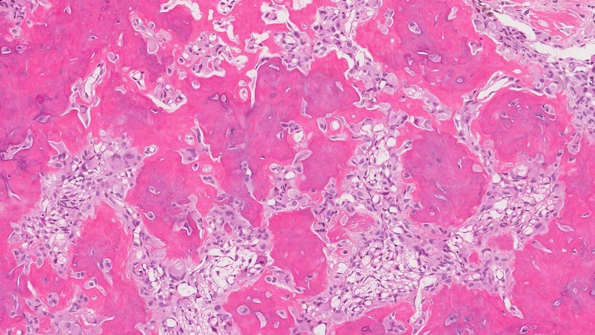
Osteosarcoma – A type of bone cancer most often seen in teenagers and young adults. It produces large amounts of abnormal osteoid. Osteosarcoma is aggressive and typically requires treatment with surgery and chemotherapy.
In the case of osteosarcoma, the ability of the tumour cells to make osteoid is one of the key features used to confirm the diagnosis.
Why is osteoid important in a pathology report?
The presence of osteoid in a tissue sample can help pathologists:
-
Confirm that the sample contains bone or bone-forming tissue.
-
Identify and classify bone tumours, including benign and malignant types.
-
Recognize conditions like heterotopic ossification that may explain symptoms or imaging findings.
Whether osteoid is a normal finding or a sign of disease depends on where it is found and what type of cells are producing it.
Questions to ask your doctor
-
-
What does the presence of osteoid mean in my report?
-
Is it a normal finding, or does it suggest a bone-forming tumour?
-
Was the osteoid associated with trauma or healing?
-
Do I need further tests or treatment based on this result?
-
Could this be related to a benign or cancerous condition?
-
We are proud to partner with:
![]()