

October 17, 2023
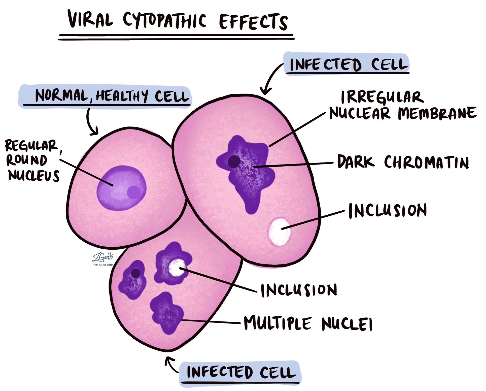
Viral cytopathic effects are changes that take place in a cell after it has been infected by a virus. These changes can only be seen after the tissue is examined under a microscope. These changes can involve the shape and size of the cell. They can also involve a part of the cell called the nucleus, which holds most of the genetic material inside a cell.
The changes that can be seen under the microscope include:
- Inclusions – These are small spots or holes inside the cytoplasm or the nucleus of the cell. The inclusions can look clear or they can have a pink color.
- Irregular nuclear membrane – The nucleus is surrounded by a thin capsule called the nuclear membrane. Normally, the membrane is smooth, but in a cell infected by a virus, it can become wrinkled.
- Chromatin changes – The genetic material inside the nucleus is called chromatin. After a cell becomes infected with a virus, the chromatin can start to look darker than normal or may move to the nuclear membrane.
- Multi-nucleated cells – Most cells have only one nucleus. Cells infected by a virus can stick together so closely that they become a single large cell. This large cell will have more than one nucleus, which pathologists call a multi-nucleated cell.
Viruses that cause viral cytopathic effects
Many different types of viruses can cause the viral cytopathic effects described above. The most common types of viruses include:
- Cytomegalovirus (CMV)
- Epstein-Barr virus (EBV)
- Human papillomavirus (HPV)
- Herpes simplex virus (HSV)
About this article
Doctors wrote this article to help you read and understand your pathology report. Contact us if you have questions about this article or your pathology report.
We are proud to partner with:
![]()
Was this article helpful?