by Jason Wasserman MD PhD FRCPC
April 8, 2024
A tubular adenoma is a type of polyp found in the large intestine which includes the colon and rectum. It starts from glandular cells that cover the inside surface of the large intestine. Tubular adenomas are considered precancerous conditions because, while most are benign (non-cancerous), they have the potential to develop into colorectal cancer called adenocarcinoma over time if left untreated. Tubular adenomas are one of the most common types of polyps found during colonoscopy screenings.
Symptoms of tubular adenomas
Most tubular adenomas do not cause symptoms and are often discovered incidentally during a routine colonoscopy or imaging study done for another reason. When symptoms do occur, they may include:
- Blood in the stool or rectal bleeding.
- Changes in bowel habits, such as constipation or diarrhea.
- Abdominal pain.
What causes a tubular adenoma?
The exact cause of tubular adenomas is not fully understood, but they are thought to result from a combination of genetic and environmental factors. Risk factors that may increase the likelihood of developing tubular adenomas include:
- Age (more common in people over 50).
- Family history of colorectal cancer or polyps.
- Certain genetic syndromes, such as familial adenomatous polyposis (FAP).
- Lifestyle factors such as diet, smoking, and lack of physical activity.
How is this diagnosis made?
The diagnosis of tubular adenoma can only be made after part or all of the adenoma is removed, and the tissue is examined under the microscope by a pathologist. The adenoma may be removed in one piece or in multiple pieces.
What does a tubular adenoma look like under the microscope?
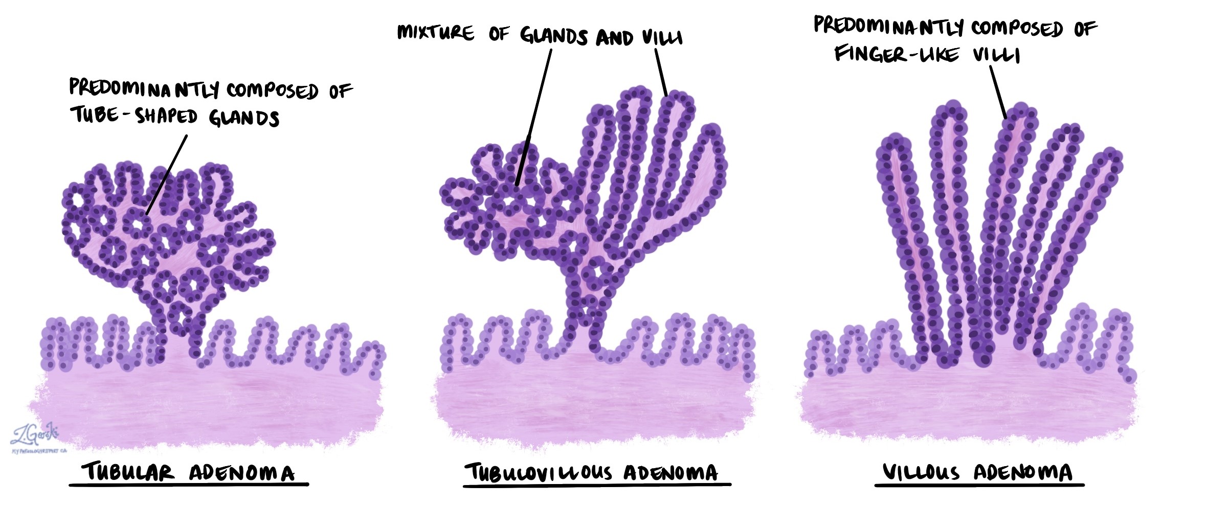
Under the microscope, a tubular adenoma of the large intestine often appears as a polyp on a stalk (pedunculated), composed of dysplastic epithelial cells forming tube-shaped glands. Compared to normal cells, these dysplastic cells exhibit increased nuclear size, hyperchromasia (increased staining intensity), and a higher nucleus-to-cytoplasm ratio. In contrast, a tubulovillous adenoma contains a mix of both tubular and finger-like villous structures, and a villous adenoma is predominantly composed of villous architecture.
Low versus high grade dysplasia in a tubular adenoma
All tubular adenomas show an abnormal pattern of growth called dysplasia. Dysplasia is important because it is a precancerous change that can become cancerous over time. When examining a tubular adenoma, pathologists divide dysplasia into two levels: low grade dysplasia and high grade dysplasia.
Tubular adenoma with low grade dysplasia
Low grade dysplasia is an early precancerous change seen in most tubular adenomas. If left untreated, low grade dysplasia can change into high grade dysplasia or cancer over time. However, the overall risk of this change developing is low.
Tubular adenoma with high grade dysplasia
High grade dysplasia is a more advanced precancerous change seen in a minority of tubular adenomas. If left untreated, tubular adenomas with high grade dysplasia can turn into a type of colon cancer called adenocarcinoma. If possible, all tubular adenomas with high grade dysplasia should be removed completely.
Margins
A margin is any tissue that the surgeon cut to remove the tubular adenoma from your body. Dysplasia at the cut edge of the tissue means that the abnormal tissue may not have been completely removed from the body.
Some tubular adenomas grow on a piece of tissue called a stalk, and the adenoma is removed by cutting the stalk. In these cases, the margin is the part of the cut stalk. However, most tubular adenomas are removed and sent to pathology as multiple pieces (fragments) of tissue. In these cases, it may not be possible for your pathologist to determine which piece is the real margin and the changes seen at the margin will not be described in your report.

Do you have a question about your pathology report?
Our team of experts is here to help.
Ask a Pathologist.
Our work is generously supported by:
![]()