Atypical Ductal Hyperplasia (ADH): Understanding Your Pathology Report
Atypical ductal hyperplasia (ADH) is a non-cancerous (benign) change in the breast that starts in the cells lining the small tubes called ducts. Although it is not cancer, ADH is considered a precancerous condition because having it is associated with an increased chance of developing breast cancer in the future. Compared with someone without ADH, a person with this condition has about a 3- to 5-fold higher chance of developing invasive ductal carcinoma over their lifetime. Even so, the overall risk from ADH alone remains modest, and most people with ADH never develop breast cancer. This article will help you understand the findings in your pathology report, what each term means, and why it matters for your care.
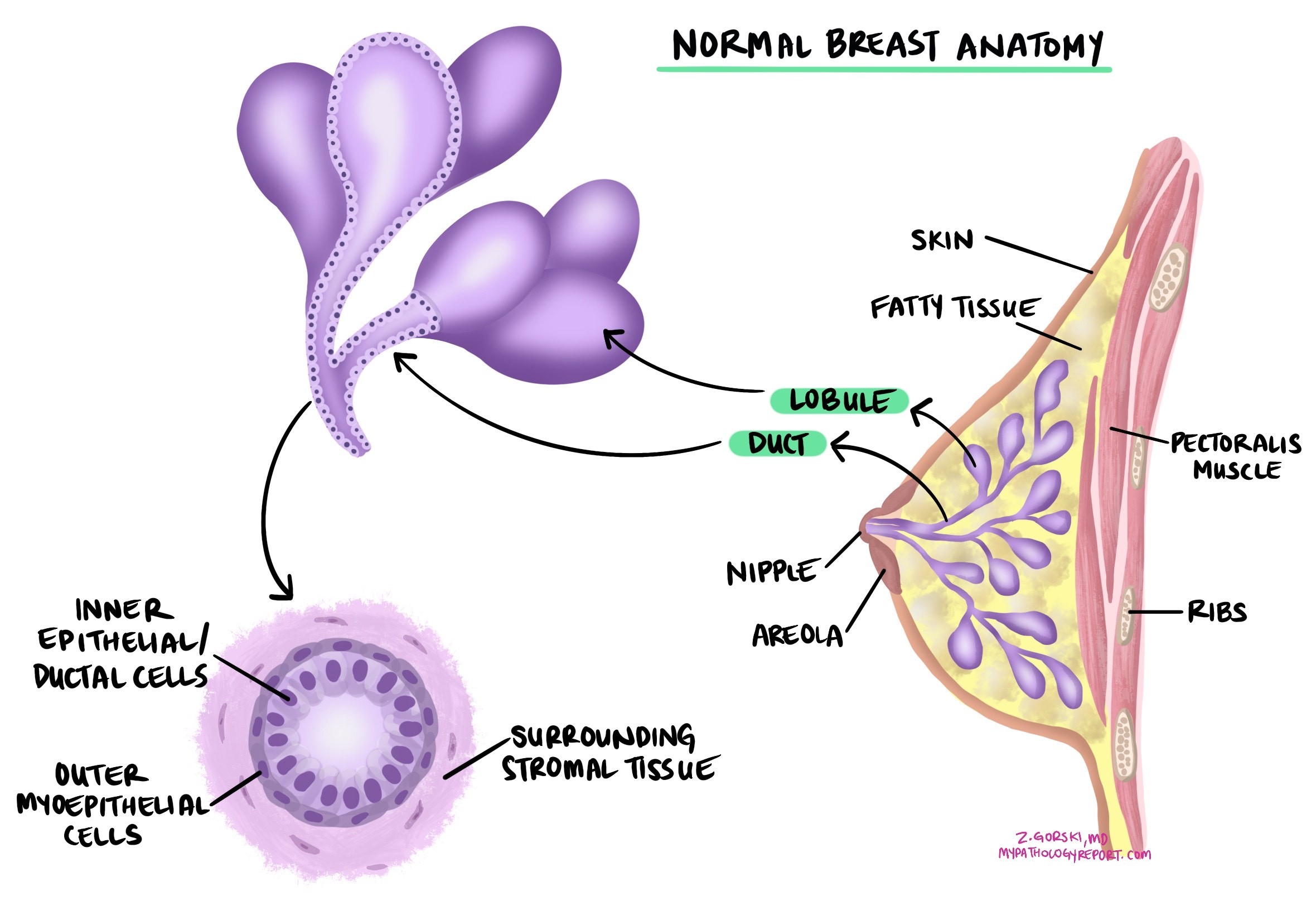
Is atypical ductal hyperplasia a type of cancer?
No. Atypical ductal hyperplasia is not breast cancer, and the abnormal cells cannot spread to other parts of the body. It is best understood as a marker that a person is at somewhat higher risk of developing breast cancer in the future. The increased risk applies to both breasts, not only the breast where ADH was found.
What causes atypical ductal hyperplasia?
Atypical ductal hyperplasia is a hormone-sensitive condition, which means the cells grow and divide in response to hormones, especially estrogen. For this reason, it is believed to be caused by the same genetic and environmental factors that lead to hormone-sensitive types of breast cancer. Lifetime estrogen exposure, family history, and increasing age all contribute to the risk.
What are the symptoms of atypical ductal hyperplasia?
Most people with atypical ductal hyperplasia have no symptoms. It usually cannot be felt as a lump. Instead, it is most often discovered when a breast imaging test, such as a mammogram, shows small calcium deposits called microcalcifications, prompting a biopsy.
How is the diagnosis made?
Doctors diagnose atypical ductal hyperplasia after removing a small sample of breast tissue in a biopsy, most often a core needle biopsy performed because of microcalcifications or another change seen on a mammogram. A pathologist examines the tissue under the microscope. In atypical ductal hyperplasia, the number of cells lining the ducts increases, and the cells appear uniform and evenly spaced (pathologists often describe them as monotonous). Microcalcifications are commonly present. On immunohistochemistry, cells are usually positive for the estrogen receptor (ER) and progesterone receptor (PR), indicating they can respond to hormones.
Because atypical ductal hyperplasia can look very similar to low-grade ductal carcinoma in situ, and because a small biopsy sample only samples part of the area, the pathologist cannot always be certain that a more advanced change is not present nearby. This is why the diagnosis from a small sample often affects what happens next, as described below.

What is the difference between atypical ductal hyperplasia and ductal carcinoma in situ?
Atypical ductal hyperplasia and ductal carcinoma in situ (DCIS) are made up of cells that look very similar under the microscope. The difference lies in amount and extent: atypical ductal hyperplasia involves only a small area and may not completely fill the ducts, whereas DCIS involves a larger area. Because the two look alike, it can be difficult for a pathologist to tell them apart from a small biopsy sample, and a larger sample may be needed to be sure which one is present.
What is the risk of developing breast cancer?
Atypical ductal hyperplasia is best thought of as a risk marker. Having it raises the lifetime chance of developing breast cancer to about 3 to 5 times that of a person without the condition, and the increased risk applies to both breasts. Despite this, the overall risk remains modest, and most people with atypical ductal hyperplasia never develop breast cancer. A strong personal or family history of breast cancer adds to the overall level of risk.
What happens after this diagnosis?
Because atypical ductal hyperplasia is a risk marker rather than a cancer, the goals after diagnosis are to make sure no more advanced change is present and to lower the future risk of breast cancer. Care is often coordinated with a breast surgeon and, when appropriate, a medical oncologist. The pathology findings guide which options the team discusses, rather than dictating a single path.
- Surgical excision — When atypical ductal hyperplasia is found on a core needle biopsy, the area is often removed with a small surgery. This is because, in a notable proportion of cases (often cited as roughly 10% to 20%), examining the whole area reveals ductal carcinoma in situ or a small invasive cancer that the needle biopsy did not capture.
- Surveillance — Regular clinical breast examinations and imaging help find any future cancer early.
- Risk-reducing medication — Because the cells are hormone-sensitive, medications that block or lower estrogen, such as tamoxifen, raloxifene, or an aromatase inhibitor, can substantially reduce the risk of developing breast cancer and may be discussed.
- Genetic counseling — Considered when there is a strong family history of breast cancer.
Questions to ask your doctor
- Was my atypical ductal hyperplasia found on a needle biopsy, and do I need surgery to remove the area?
- If the area is removed, will you be checking for ductal carcinoma in situ or invasive cancer?
- How much does this finding increase my risk of developing breast cancer?
- Does the increased risk apply to both breasts?
- Were the cells positive for the estrogen and progesterone receptors?
- What surveillance schedule and imaging do you recommend?
- Would a risk-reducing medication such as tamoxifen or an aromatase inhibitor be appropriate for me?
- Should I be referred for genetic counseling based on my personal or family history?
- Were any other breast changes found in the same sample?
- What signs or symptoms should prompt me to contact you between visits?
Related articles on MyPathologyReport.com
- Atypical Lobular Hyperplasia of the Breast: Understanding Your Pathology Report
- Ductal Carcinoma In Situ (DCIS) of the Breast: Understanding Your Pathology Report
- Lobular Carcinoma In Situ (LCIS): Understanding Your Pathology Report
- Invasive Ductal Carcinoma of the Breast: Understanding Your Pathology Report
- Understanding Your Breast Biopsy Pathology Report