Immunohistochemistry (IHC) is a widely used laboratory test that involves the use of antibodies to detect specific antigens (proteins) in cells within tissue sections. Pathologists use this test to see the distribution and localization of specific proteins within different parts of a tissue, thereby providing valuable diagnostic, prognostic, and predictive information.
How does immunohistochemistry work?
The principle behind immunohistochemistry is based on the specific binding affinity between an antibody and its antigen. The antibody is designed to target and bind to a specific protein of interest within the tissue sample. Once bound, this interaction is visualized using a detection system, resulting in a colored or fluorescent signal that can be seen under a microscope.
Steps involved in immunohistochemistry
- Sample preparation: Tissue specimens are collected, often through biopsy or surgical resection, and then fixed to preserve tissue architecture. Formalin is a commonly used fixative. The tissue is embedded in paraffin wax to facilitate sectioning.
- Sectioning: The paraffin-embedded tissue block is cut into thin sections (usually 4-5 micrometers thick) using a microtome. These sections are placed on microscope slides for staining.
- Deparaffinization and rehydration: The slides are treated to remove the paraffin and rehydrate the tissues, typically using xylene (or alternatives) followed by graded alcohols.
- Antigen retrieval: Many antigens become masked during the fixation process. Antigen retrieval involves treating the sections with heat or enzymes to expose these antigenic sites, making them accessible to antibodies.
- Blocking: Non-specific binding sites are blocked using a protein solution to prevent the primary antibody from binding nonspecifically, which could lead to false-positive results.
- Primary antibody incubation: The slide is incubated with a primary antibody that is specific to the antigen of interest. This step allows the antibody to bind to its target antigen in the tissue.
- Detection: After washing away any unbound primary antibody, a secondary antibody is added. This antibody is conjugated to an enzyme (like horseradish peroxidase or alkaline phosphatase) or a fluorescent label and is designed to bind to the primary antibody. The presence of the secondary antibody is then visualized through a colorimetric reaction (in the case of enzyme-conjugated antibodies) or fluorescence (in the case of fluorescently labeled antibodies). For colorimetric detection, a substrate is added that the enzyme converts into a visible, colored product at the site of the antigen-antibody interaction.
- Counterstaining: To enhance visualization of the tissue architecture, a mild counterstain (e.g., hematoxylin) is typically applied to the slide, staining cell nuclei with a contrasting color.
- Mounting and visualization: The slide is covered with a coverslip, and the stained tissue is examined under a light or fluorescent microscope. The localization, intensity, and pattern of staining provide insights into the presence and distribution of the antigen within the tissue.
Applications
Immunohistochemistry is instrumental in diagnostic pathology for identifying the type and origin of cancer cells, diagnosing infectious diseases, and differentiating between similar-looking conditions.
Through its ability to specifically identify proteins within the complex architecture of tissues, immunohistochemistry has become an indispensable tool in pathology, significantly impacting diagnostics, prognostics, and the development of targeted therapies.
Patterns of expression in immunohistochemistry
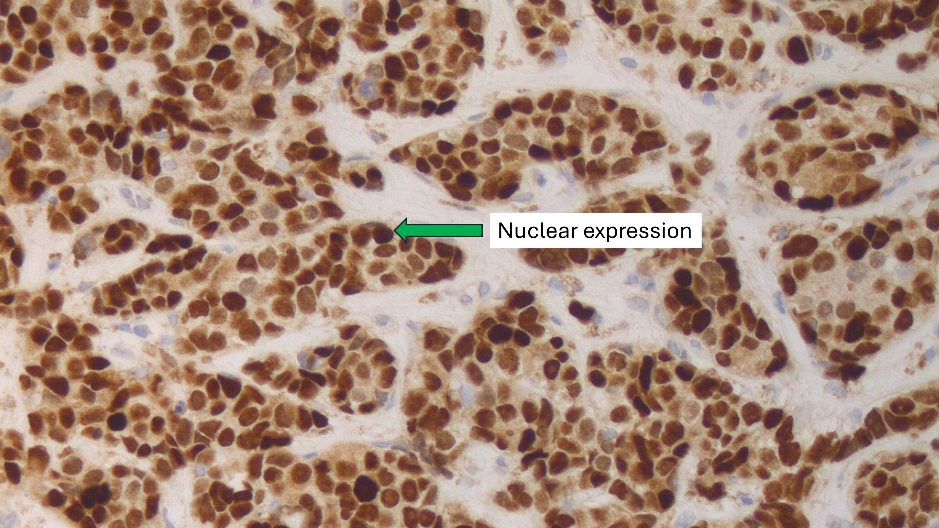
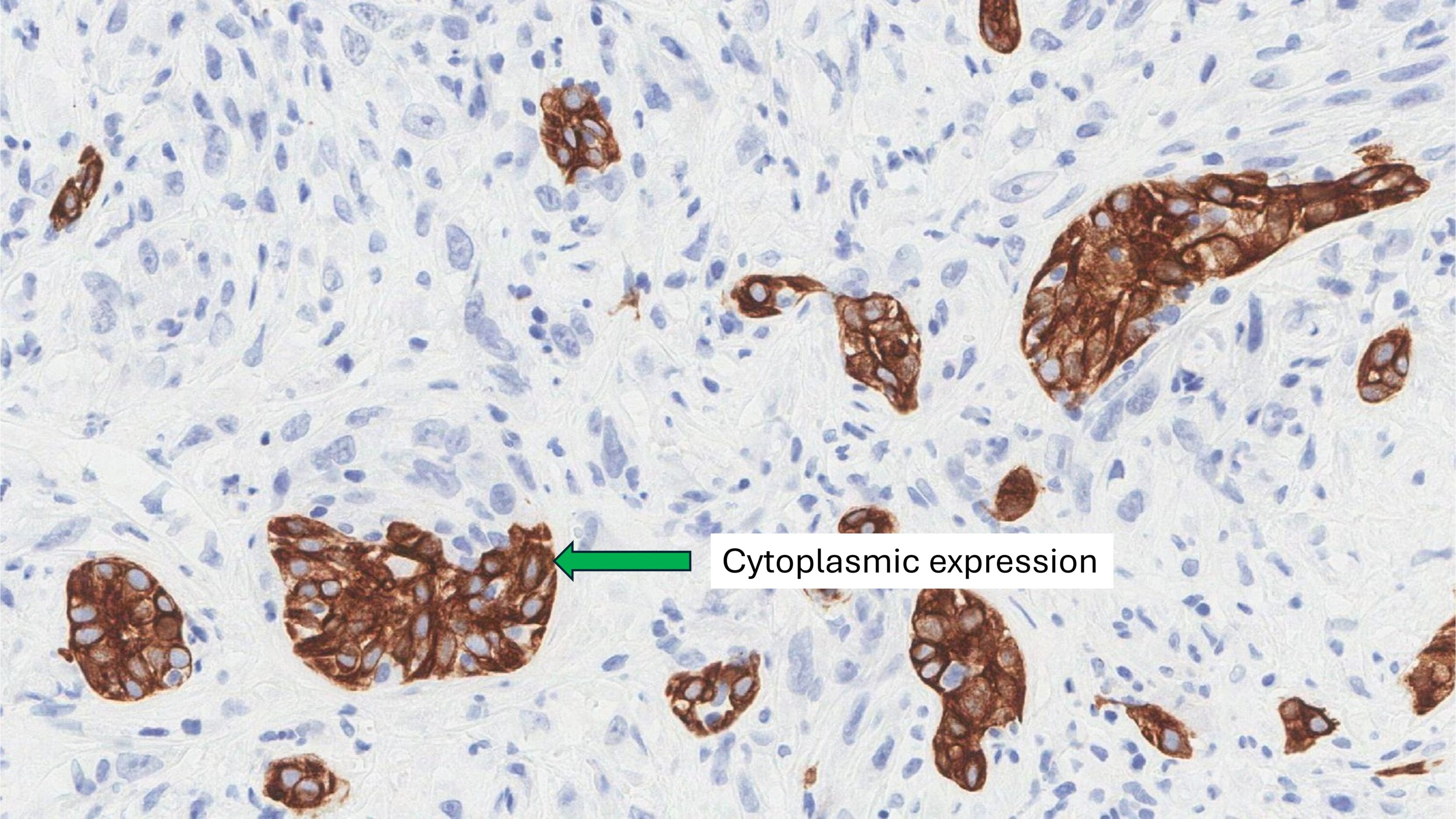
In immunohistochemistry, the patterns of staining—nuclear, cytoplasmic, and membranous—refer to the localization of the antigen (protein) within different compartments of the cell. Each pattern provides valuable insights into the function of the protein and the type of cell expressing the protein.
Nuclear expression
Nuclear expression occurs when the IHC staining is localized to the cell nucleus, where DNA and RNA synthesis occur, and many regulatory proteins are located. Examples of proteins that show nuclear expression include transcription factors, nuclear receptors, and proteins involved in DNA replication and repair. For instance, the estrogen receptor (ER) in breast cancer cells shows nuclear staining because it acts as a transcription factor that regulates gene expression.

Nuclear staining is significant in diagnosing diseases that involve alterations in gene expression or cell cycle regulation. It is particularly important in cancers where the presence or absence of nuclear proteins, such as hormone receptors, can guide treatment decisions.
Cytoplasmic Expression
Cytoplasmic expression is observed when the staining is distributed throughout the cytoplasm, the part of the cell that surrounds the nucleus and contains various organelles and the cytoskeleton.
Examples of proteins that show cytoplasmic expression include enzymes, structural proteins, and certain signaling molecules. An example includes cytokeratins, which are intermediate filament proteins found in the cytoplasm of epithelial cells.

Cytoplasmic staining helps identify cells that are producing specific proteins involved in metabolism, signaling, or cellular structure. This information can be crucial for diagnosing and classifying tumors, understanding metabolic diseases, and identifying infectious agents.
Membranous Expression
Membranous expression refers to staining that is localized to the cell membrane, the boundary that separates the cell from its external environment and mediates communication with other cells and the extracellular matrix. Examples of proteins that show membranous expression include membrane receptors, transporters, and cell adhesion molecules. A well-known example is HER2/neu overexpression in certain breast cancers, where HER2 protein is detected as a membranous staining pattern.

Membranous staining is particularly important for identifying cells responding to extracellular signals or involved in cell-cell or cell-matrix interactions. In oncology, the presence of specific membrane proteins can indicate the aggressiveness of a tumor and its susceptibility to targeted therapies.
Understanding these patterns of expression is fundamental in the application of immunohistochemistry in diagnostic pathology. It enables pathologists to make precise diagnoses, understand the pathophysiology of diseases, and inform treatment strategies. For example, determining the presence of ER (nuclear expression) and HER2 (membranous expression) in breast cancer cells is crucial for deciding on hormone therapy and targeted therapy, respectively.
Common immunohistochemical markers
CD34
Cytokeratin 7 (CK7)
Cytokeratin 20 (CK20)
Desmin
Estrogen receptor (ER)
GATA-3
Ki-67
MIB-1
p16
p63
p53
p40
Progesterone receptor (PR)
S100
SOX-10
TTF-1
About this article
Doctors wrote this article to help you read and understand your pathology report. Contact us if you have questions about this article or your pathology report. For a complete introduction to your pathology report, read this article.

