Mucinous Adenocarcinoma of the Lung: Understanding Your Pathology Report
Mucinous adenocarcinoma is a type of lung cancer and one of the main subtypes of non-small cell lung cancer. This tumour gets its name because the cancer cells produce large amounts of a sticky fluid called mucin. Mucinous adenocarcinoma typically develops in the outer parts of the lungs, and it can sometimes affect multiple areas or even both lungs at the same time.
What causes mucinous adenocarcinoma of the lung?
The most common cause of mucinous adenocarcinoma is tobacco smoking. People who smoke cigarettes or have smoked in the past have a higher chance of developing this tumour. Other causes, although less common, include exposure to harmful substances such as radon gas, air pollution, or workplace chemicals.
Mucinous adenocarcinoma often exhibits genetic changes involving the KRAS gene. These genetic changes help tumour cells grow and survive. Other genetic alterations include changes in genes called NRG1, ALK, and ROS1.
What are the symptoms of mucinous adenocarcinoma of the lung?
Symptoms of mucinous adenocarcinoma may include:
-
Persistent cough.
-
Coughing up mucus or fluid (called bronchorrhea).
-
Shortness of breath or difficulty breathing.
-
Chest pain or discomfort.
-
Unexplained weight loss.
-
Fatigue.
On imaging studies such as CT scans, this tumour may look like pneumonia, causing doctors to initially suspect an infection rather than cancer.
How is mucinous adenocarcinoma diagnosed?
The diagnosis of mucinous adenocarcinoma is usually made after a small sample of tissue is removed from the lung in a procedure called a biopsy. A biopsy can be performed using a needle or during a surgery. Once removed, the sample is sent to a pathologist who examines it closely under a microscope to confirm the diagnosis.
What does mucinous adenocarcinoma look like under the microscope?
Under the microscope, mucinous adenocarcinoma is composed of tumour cells arranged in glandular or columnar patterns. These cells produce abundant mucin, giving the tumour a distinctive jelly-like or mucinous appearance. The nuclei (the part of the cell containing genetic material) are small and usually located at the base of the cell. Pathologists refer to these cells as “columnar” or “goblet” cells due to their distinctive shape.
The tumour often grows along the inner surfaces of the small air sacs (alveoli) of the lungs. This pattern of growth is called “lepidic growth.” However, the tumour can also invade deeper into the lung tissue using other growth patterns, such as solid or papillary.
What other tests are done to confirm the diagnosis?
Your pathologist may perform special tests to help confirm the diagnosis of mucinous adenocarcinoma and to rule out cancers that have spread from other parts of the body. One important test is called immunohistochemistry. In this test, special markers are used to identify proteins made by the tumour cells.
The typical results of immunohistochemistry for mucinous adenocarcinoma include:
-
CK7 – Usually positive.
-
CK20 – Sometimes positive.
-
CDX2 – Sometimes positive.
-
TTF-1 – Usually negative.
-
Napsin A – Usually negative.
Your pathologist may also perform genetic testing (molecular testing) to look for genetic changes commonly found in mucinous adenocarcinoma. These tests look for alterations in genes such as KRAS, NRG1, ALK, and ROS1, which can be important for guiding treatment.
Can mucinous adenocarcinoma spread to other parts of the body?
Yes. Although mucinous adenocarcinoma tends to grow relatively slowly compared to other types of lung cancer, it can still metastasize to other parts of the body. It frequently spreads within the lungs through airways (a process called aerogenous spread), which can result in tumour growth in multiple areas of the lungs. It can also spread to lymph nodes and distant sites such as the bones, liver, and brain.
Spread through air spaces
Spread through air spaces (STAS) describes a pattern of invasion observed in lung cancer, where cancer cells are seen spreading into the air spaces in the lung tissue outside the tumour. The presence of STAS has been associated with a higher risk of recurrence and worse overall survival in patients with adenocarcinoma of the lungs, especially in those with early-stage disease. Recognizing STAS can therefore provide valuable prognostic information and help in risk stratification.
Pathologists identify STAS by carefully examining the lung tissue surrounding the tumour under a microscope. They look for tumour cells or clusters of cells within the air spaces that are separate from the main tumour and not attached to the tumour edge, often located at a distance from the tumour mass itself. These cells can be free-floating or attached to the alveolar walls, but are distinguishable from the primary tumour and not explained by other processes such as artefact or lymphovascular invasion.
Multiple tumours
It is not uncommon for more than one tumour to be found in the same lung. When this happens, each tumour will be described separately in your report.
There are two possible explanations for finding more than one tumour:
- The tumour cells from one tumour have spread to another part of the lung. This explanation is more likely when all of the tumours are of the same histologic type. For example, if all of the tumours are acinar-type adenocarcinoma. If the tumours are on the same side of the body, the smaller tumours are called nodules. If the tumours are on different sides of the body (right and left lung), the smaller tumour is called metastasis.
- The tumours have developed separately. This is the more likely explanation when the tumours are of different histologic types. For example, one tumour is an adenocarcinoma while the other is a squamous cell carcinoma. In this situation, the tumours are considered separate primaries and not metastatic disease.
Pleural invasion
The pleura is a thin layer of tissue that covers the lungs and lines the inner surface of the chest cavity.
It has two layers:
-
Visceral pleura: The layer attached directly to your lungs.
-
Parietal pleura: The layer lining the chest wall and diaphragm.
When tumour cells grow beyond the lung and invade the pleura, it is called pleural invasion. Pleural invasion is important because it affects both staging and prognosis:
-
Tumour stage: Tumours invading the pleura are considered more advanced. Pleural invasion increases the tumour’s T-stage in the TNM staging system.
-
Prognosis: Patients with pleural invasion generally have a worse prognosis because the cancer is more aggressive and likely to spread.
Lymphovascular invasion
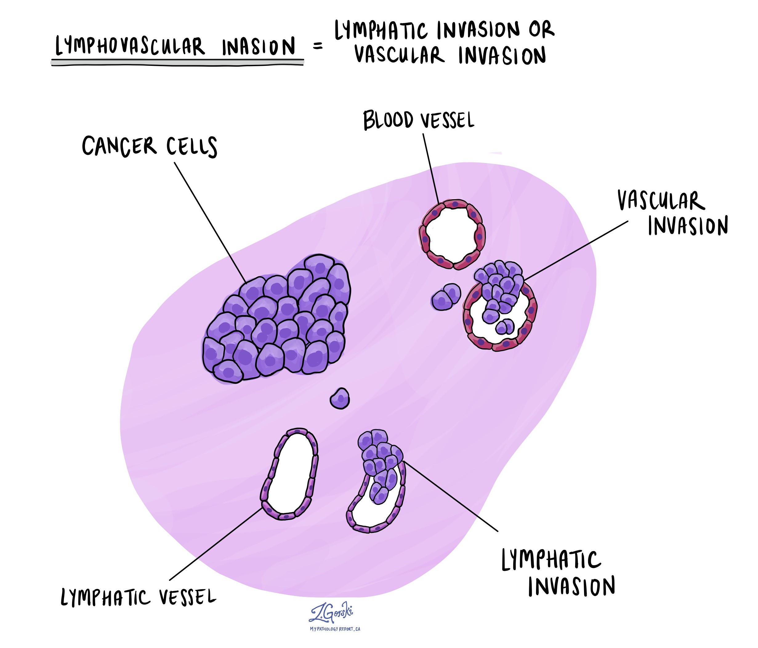
Cancer cells can spread into tiny blood vessels or lymphatic channels, a process called lymphovascular invasion. Blood vessels carry blood throughout the body, while lymphatic channels carry lymph fluid, which plays a crucial role in immune function. When tumour cells enter these channels, they can spread to other parts of the body, such as lymph nodes, the liver, or bones. Finding lymphovascular invasion means a higher risk of cancer spreading.
Margins
In pathology, a margin refers to the edge of tissue removed during surgery to remove a tumour. After lung surgery, pathologists carefully examine all of these tissue edges under a microscope to determine if the tumour has been completely removed.
Margins assessed in lung cancer surgeries typically include:
-
Bronchial margin – This is where the surgeon cuts through the airway.
-
Vascular margin – These are the areas where large blood vessels near the tumour are cut.
-
Parenchymal margin – This margin includes the edge of lung tissue around the tumour.
-
Pleural margin – The pleura is a thin lining surrounding the lung, and this margin is examined to determine if the tumour grows close to or through this lining.
Margins can be described in two ways:
-
Negative margin – No cancer cells are seen at any cut edge. This indicates that the tumour has likely been entirely removed, which is the goal of surgery.
-
Positive margin – Cancer cells are seen at the cut edge of the tissue. A positive margin means there could still be tumour cells remaining in your body. Patients with a positive margin may require additional treatments, such as a second surgery or radiation therapy, to remove any remaining tumour cells and reduce the risk of recurrence.
The status of the margins helps your doctor determine the need for additional treatment and plays an important role in predicting the likelihood of the tumour growing back.

Lymph nodes
Lymph nodes are small, bean-shaped organs that play an essential role in the immune system. They are connected throughout the body by small channels called lymphatic vessels. Cancer cells can spread from a tumour through these lymphatic vessels and into nearby lymph nodes—a process called lymph node metastasis.
Lymph nodes in the lungs and chest are grouped into specific areas, known as lymph node stations. There are 14 different lymph node stations, each with a specific location:
-
Station 1: Lower cervical, supraclavicular, and sternal notch lymph nodes.
-
Station 2: Upper paratracheal lymph nodes.
-
Station 3: Prevascular and retrotracheal lymph nodes.
-
Station 4: Lower paratracheal lymph nodes.
-
Station 5: Subaortic (aortopulmonary window) lymph nodes.
-
Station 6: Para-aortic lymph nodes (near the ascending aorta or phrenic nerve).
-
Station 7: Subcarinal lymph nodes (below the carina, where the trachea splits into bronchi).
-
Station 8: Paraesophageal lymph nodes (alongside the esophagus below the carina).
-
Station 9: Pulmonary ligament lymph nodes.
-
Station 10: Hilar lymph nodes (at the lung hilum, where airways enter the lung).
-
Station 11: Interlobar lymph nodes (between lung lobes).
-
Station 12: Lobar lymph nodes (within lung lobes).
-
Station 13: Segmental lymph nodes (within lung segments).
-
Station 14: Subsegmental lymph nodes (within smaller lung subsegments).

If lymph nodes are removed during surgery, a pathologist carefully examines them under a microscope to see if they contain cancer cells. The pathology report typically includes:
-
The total number of lymph nodes examined.
-
The locations (stations) of the lymph nodes examined.
-
The number of lymph nodes containing cancer cells.
-
The size of the largest group of cancer cells (often called a “focus” or “deposit”).
Lymph node examination provides important information that helps your doctor determine the cancer’s pathologic nodal stage (pN). It also helps predict the likelihood that cancer cells may have spread to other parts of the body, guiding decisions about additional treatments such as chemotherapy, radiation therapy, or immunotherapy.
How is mucinous adenocarcinoma of the lung staged?
Your doctor uses the TNM staging system to describe how advanced your tumour is. This system uses information about the tumour size and spread (T), lymph node involvement (N), and the presence of cancer cells in distant parts of the body (M).
Tumour size and spread (T-stage)
-
T1: The tumour is no larger than 3 cm and has not spread beyond the lung.
-
T2: The tumour is between 3 and 5 cm, or has grown into the lining of the lung (pleura), or blocks a large airway.
-
T3: The tumour is between 5 and 7 cm, or has invaded into the chest wall, diaphragm, or the outer lining of the heart (pericardium).
-
T4: The tumour is larger than 7 cm, involves nearby vital structures (e.g., the heart or major blood vessels), or there are multiple tumours within the same lung.
Lymph node involvement (N-stage)
-
NX: Lymph nodes were not examined.
-
N0: No cancer cells were found in the examined lymph nodes.
-
N1: Cancer cells were found in lymph nodes inside or very close to the lung.
-
N2: Cancer cells were found in lymph nodes around the large airways or in the central chest (mediastinum).
-
N3: Cancer cells were found in lymph nodes on the opposite side of the chest or in the neck region.
Metastatic spread (M-stage)
-
M0: No spread of cancer cells to distant organs.
-
M1: Cancer cells have spread to distant parts of the body, such as the opposite lung, brain, bones, or liver.
A higher stage (T, N, or M) means the cancer is more advanced and usually has a worse prognosis.
What is the prognosis for mucinous adenocarcinoma of the lung?
The prognosis (expected outcome) for mucinous adenocarcinoma can vary. It depends on several factors, including tumour stage, lymph node involvement, genetic changes, and whether the tumour has spread to other parts of the body.
Historically, mucinous adenocarcinoma was thought to have a poorer prognosis compared to non-mucinous adenocarcinoma. However, recent studies have suggested that the outcome can be similar, especially when the disease is diagnosed at an early stage. Patients with tumours limited to the lung without lymph node involvement typically have a better prognosis than those with advanced disease.
Biomarkers for mucinous adenocarcinoma of the lung
Biomarkers are specific molecules found inside tumour cells. These molecules help doctors understand how the tumour behaves and how it might respond to different treatments. Testing for biomarkers is important in lung cancer because some tumours contain genetic changes or alterations that make them respond well to targeted therapies. Targeted therapies are drugs designed specifically to attack cancer cells with these genetic changes. Identifying these biomarkers helps doctors choose the most effective treatment options.
Pathologists look for biomarkers using specialized laboratory tests. Two common tests include:
-
Next-generation sequencing (NGS) – This test examines many genes at the same time to find mutations (changes in the genetic material of tumour cells). NGS can quickly identify multiple biomarkers from a single tissue sample.
-
Immunohistochemistry (IHC) – This test uses special stains that attach to specific proteins produced by cancer cells. When these proteins are present, the tumour cells change colour under the microscope. IHC helps confirm whether certain biomarkers are present in the tumour.
Common biomarkers tested in mucinous adenocarcinoma of the lung
Your pathology report may include information about the following biomarkers. Each biomarker can help guide your treatment and provide important information about your tumour.
-
EGFR: Mutations (changes) in the EGFR gene are common in lung adenocarcinoma, particularly in individuals who have never smoked, women, and those of East Asian ancestry. Tumours with EGFR mutations often respond very well to targeted therapies called EGFR inhibitors. Your report will describe the tumour as EGFR-positive if a mutation is found. If no mutation is found, it will be called EGFR-negative.
-
ALK: Changes in the ALK gene, known as ALK rearrangements or fusions, lead to tumour growth and are often found in younger patients or non-smokers. Tumours with ALK gene rearrangements usually respond well to medications called ALK inhibitors. Your report will say your tumour is ALK-positive if this change is present. If it is not present, your tumour will be ALK-negative.
-
ROS1: ROS1 rearrangements (fusions) cause cancer cells to grow quickly. ROS1-positive tumours typically respond well to targeted ROS1 inhibitor therapies. If your tumour has a ROS1 rearrangement, it will be described as ROS1-positive. If no rearrangement is found, it will be described as ROS1-negative.
-
BRAF: Certain mutations in the BRAF gene can cause tumour cells to grow rapidly. Tumours with specific BRAF mutations, particularly the V600E mutation, can be treated with BRAF inhibitors. If a BRAF mutation is found, your tumour will be described as BRAF-positive. If no mutation is found, it will be called BRAF-negative.
-
MET: Mutations in the MET gene, especially mutations leading to “MET exon 14 skipping,” cause increased tumour growth. MET-positive tumours often respond to targeted therapies known as MET inhibitors. Your pathology report will describe your tumour as MET-positive if this mutation is present. If no mutation is found, your tumour will be MET-negative.
-
RET: RET rearrangements or fusions result in uncontrolled tumour growth. Tumours with RET fusions usually respond very well to RET inhibitors. Your report will state that your tumour is RET-positive if a fusion is found. If a fusion is not found, it will be described as RET-negative.
-
NTRK1-3: NTRK gene fusions are rare but can strongly promote tumour growth. Tumours with NTRK fusions usually respond to targeted medications known as TRK inhibitors. If an NTRK fusion is detected, your tumour will be described as NTRK-positive. If not, it will be described as NTRK-negative.
-
KRAS: KRAS mutations are common in mucinous adenocarcinomas, especially among smokers. Historically, KRAS-positive tumours were difficult to treat, but recent drugs targeting a specific KRAS mutation (KRAS G12C) have shown promising results. If a KRAS mutation is present, your tumour will be described as KRAS-positive. If no mutation is detected, your tumour is KRAS-negative.
-
ERBB2 (HER2): ERBB2 mutations (also known as HER2 mutations) can drive tumour growth, particularly in non-smokers. Tumours with HER2 mutations may respond to targeted therapies currently under investigation or available in specialized centres. If your tumour has an ERBB2 mutation, it will be described as ERBB2-positive. If no mutation is found, it will be ERBB2-negative.
-
NRAS: Mutations in the NRAS gene occur most commonly in tumours of people who have smoked. Currently, targeted therapies specific to NRAS mutations are limited; however, identifying this mutation can still aid in understanding tumour behaviour. Your tumour will be described as NRAS-positive if a mutation is found or NRAS-negative if no mutation is present.
-
MAP2K1 (MEK1): MAP2K1 mutations are more common among smokers and are associated with increased tumour growth. Currently, treatments specifically targeting MAP2K1 mutations are still being studied. Your pathology report will indicate if a MAP2K1 mutation is present (MAP2K1-positive) or not (MAP2K1-negative).
-
NRG1: NRG1 gene rearrangements are rare but significant because they can promote rapid tumour growth. Researchers are actively investigating targeted treatments for tumours with NRG1 rearrangements. Your tumour will be described as NRG1-positive if this rearrangement is found. If it is not found, it will be NRG1-negative.
Why are biomarker tests important for treatment?
Identifying these biomarkers in your tumour is essential because they help doctors choose the most effective treatments. Some biomarkers match specific drugs that directly target tumour cells. These treatments often work better and have fewer side effects than traditional chemotherapy.
If your tumour does not have biomarkers that match available targeted treatments, your doctor may recommend other options, such as chemotherapy or immunotherapy. Your medical team will help you understand your test results and the best treatment options available for you.
Questions to ask your doctor
-
What stage is my mucinous adenocarcinoma?
-
Were the margins negative or positive?
-
Were any lymph nodes involved by tumour cells?
-
Are additional treatments, such as chemotherapy, targeted therapy, or radiation therapy, needed?
-
Should I undergo genetic testing of my tumour?
-
Do my genetic test results influence the choice of treatment?
-
What is my prognosis given the features of my tumour?
-
How frequently will I need follow-up appointments and scans?
-
Are there clinical trials available for my type of lung cancer?
-
What resources are available for emotional and practical support for me and my family?