Atypical: Definition
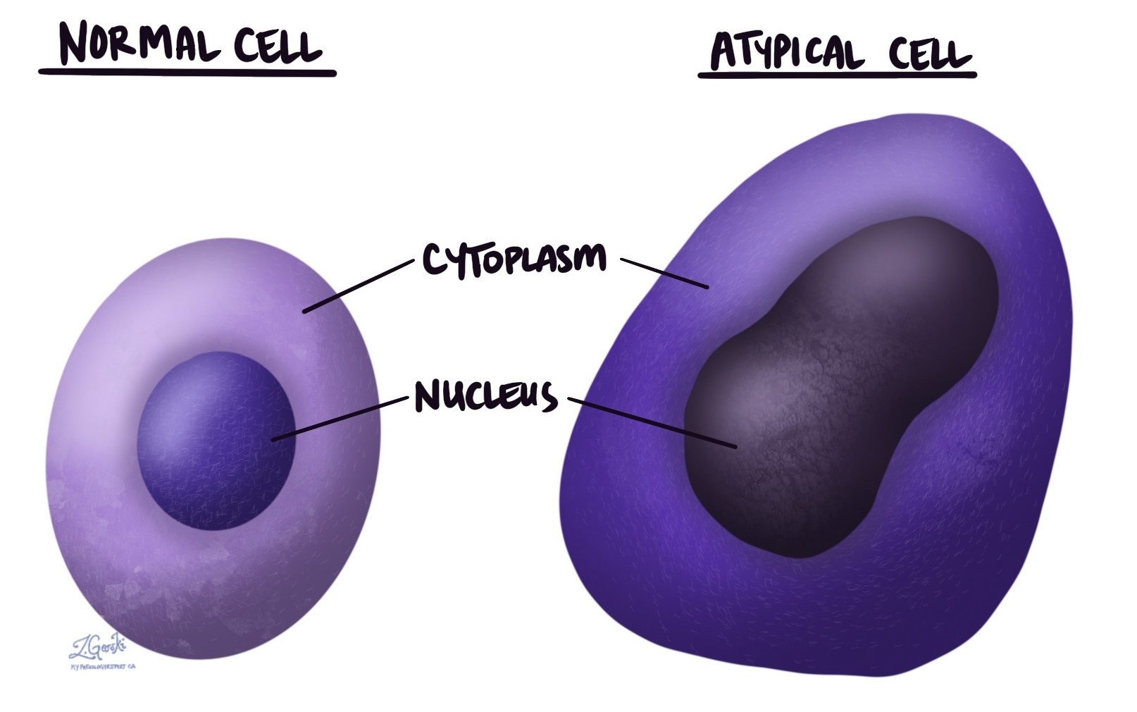
In pathology, the word “atypical” describes cells that look unusual or abnormal under the microscope. It refers to changes in cell size, shape, or internal structure, especially in the cytoplasm (the main body of the cell) or the nucleus (the part that holds the cell’s genetic material). Atypical is a description, not a diagnosis on its own. This article explains what atypical means, what can cause it, and why it may appear in your pathology report.
Does atypical mean cancer?
Not necessarily. Atypical refers to how cells look, not to what is causing the change, so it does not, by itself, mean cancer. Malignant (cancerous) tumors almost always contain atypical cells, but atypical cells also appear in many benign (noncancerous) conditions, such as inflammation or infection, and in precancerous changes. Because the same word can point to harmless changes or to something more serious, a pathologist interprets atypical cells in context, considering the rest of the tissue sample, other test results, and your medical history. In some cases, your care team may recommend additional tests or follow-up to clarify what the atypical cells mean for you.
What causes cells to become atypical?
Cells can look atypical for many different reasons, ranging from temporary and harmless to serious. Common causes include:
- Inflammation — Inflammation is the body’s natural response to injury, infection, or disease. Cells in an area of inflammation may appear atypical for a time and usually return to normal once the inflammation settles.
- Infection — Cells infected by viruses often look atypical under the microscope. Pathologists call these changes viral cytopathic effects, and the cells usually return to normal after the infection clears.
- Radiation — Radiation therapy used to treat cancer can make nearby healthy cells look atypical. These changes can last a long time, so it helps to tell your doctor and pathologist if you have had radiation.
- Precancerous changes — Conditions such as dysplasia and carcinoma in situ contain atypical cells that could develop into cancer if they are not treated. Recognizing them early allows for monitoring or treatment before cancer develops.
- Cancer — Cancer cells almost always look atypical compared with normal cells. How atypical the cells appear helps the pathologist diagnose cancer and assign its grade, which describes how abnormal the cells look and helps guide treatment decisions.
What do atypical cells look like under the microscope?
Under the microscope, atypical cells often have irregular shapes and sizes. The nucleus may be enlarged or unusually shaped, and the genetic material inside it may look darker or more disorganized than normal. Cell borders may appear indistinct, and atypical cells may stand out from the surrounding normal cells. These features help the pathologist recognize atypical cells and investigate what is causing them.
Questions to ask your doctor
- What does “atypical” mean in my pathology report?
- Were the atypical cells found in a benign, precancerous, or cancerous condition?
- Could inflammation, infection, or previous radiation cause the atypical changes?
- Do I need any additional tests or a repeat biopsy to understand the atypical cells?
- Do these atypical cells need monitoring over time?
- If this turns out to be precancerous, what are my options for monitoring or treatment?
- Based on my overall results, how concerned should I be about these atypical cells?