Basal Cells: Definition
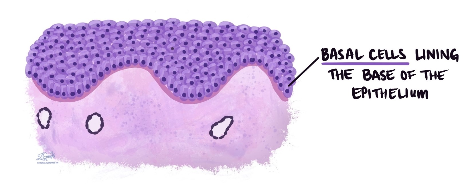
Basal cells are a type of cell found at the very bottom (the “base”) of certain tissues, such as the skin and the lining of some internal organs. They are located in a thin layer at the bottom of the epithelium just above the basement membrane. Basal cells act like stem cells because they divide to replace older cells as they are shed or damaged.
Where are basal cells found?
Basal cells are normally seen in:
-
The skin: In the epidermis, basal cells continuously divide to create new keratinocytes, which then move upward to form the protective outer surface.
-
The lining of organs: In areas like the esophagus, cervix, prostate, and urinary tract, basal cells form the lower layer of the lining and help maintain healthy tissue.
-
The respiratory tract: In the airways, basal cells help repair and regenerate the lining after injury.
Why are basal cells important?
Basal cells are important because they keep tissues healthy by replacing worn-out or injured cells. They also provide structural support to the tissue lining. Since they can divide and make new cells, basal cells are often the starting point for certain types of cancer. For example, basal cell carcinoma is the most common form of skin cancer and begins in basal cells of the skin. In the prostate, the presence or absence of basal cells helps pathologists distinguish between benign (non-cancerous) and malignant (cancerous) conditions.
What do basal cells look like under the microscope?
Under the microscope, basal cells usually appear as small, darker-staining cells that form a continuous layer along the basement membrane. They often have oval or round nuclei and a limited amount of cytoplasm. In healthy tissue, basal cells are neatly arranged in a single layer at the base of the epithelium.
What does it mean if basal cells are mentioned in my pathology report?
If your pathology report mentions basal cells, the pathologist is likely describing how this layer of cells appears under the microscope and whether it looks normal or abnormal. For example:
-
In the skin, basal cells may look normal, enlarged, or show signs of cancer.
-
In the cervix, basal cells may be increased in number in precancerous conditions.
-
In the prostate, basal cells are usually present in non-cancerous tissue but are often absent in prostate cancer.
Pathologists may also use special tests, such as immunohistochemistry, to highlight basal cells and confirm their presence or absence.
Conditions associated with basal cells
-
Normal tissue repair: Basal cells divide to replace damaged cells in the skin, cervix, or other tissues.
-
Basal cell carcinoma: A common skin cancer that starts in basal cells of the epidermis.
-
Precancerous changes: An increase in basal cells can sometimes be seen in conditions such as cervical intraepithelial neoplasia or Barrett’s esophagus.
-
Prostate cancer diagnosis: The absence of basal cells in glands of the prostate is one of the features pathologists use to confirm cancer.
-
Basaloid neoplasms: Growths made up of basal-like cells that can be either benign or malignant.
Questions to ask your doctor
-
Why are basal cells mentioned in my report?
-
Do the basal cells in my sample look normal or abnormal?
-
Does the presence or absence of basal cells affect my diagnosis?
-
Were any special tests performed to look for basal cells?
-
How does this finding guide my treatment or follow-up care?