Viral Cytopathic Effects: Definition
Viral cytopathic effects are changes that appear in a cell after a virus infects it. “Cytopathic” means harmful to cells. A pathologist sees these changes under the microscope. They can alter a cell’s size and shape and change the appearance of its nucleus.
Viral cytopathic effects are a finding, not a diagnosis. They show that a virus has probably infected cells in your sample. This article explains what these changes look like, which viruses cause them, how pathologists identify them, and what your report wording means.
Why do viruses change cells?
A virus changes a cell because it uses the cell to make copies of itself. The virus enters the cell and takes over its machinery. As copies build up, the cell’s structure changes. Different viruses cause different changes. A skilled pathologist can often suspect the virus from the pattern.
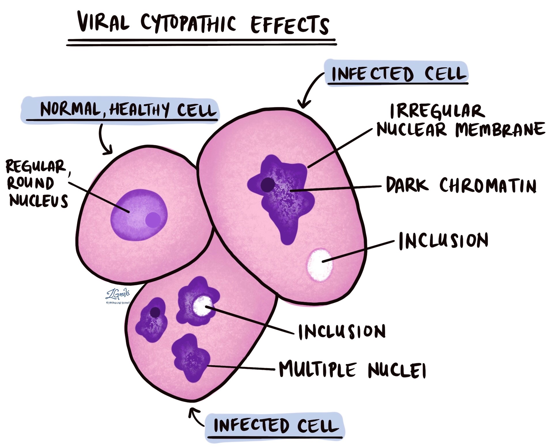
What changes do pathologists see?
Viral cytopathic effects can involve any part of the cell. The most common changes are:
- Inclusions. These are spots or clear areas inside the nucleus or the cytoplasm. They may look pink or clear. They often contain viral particles. See intranuclear inclusions.
- Enlarged nucleus. The nucleus can swell. In cytomegalovirus infection, it can become very large with a clear area around the inclusion.
- Ground-glass nucleus. The nucleus looks smooth and clear, like frosted glass. The chromatin, the cell’s genetic material, moves to the nuclear edge. Pathologists call this margination.
- Irregular nuclear shape. The normally smooth edge of the nucleus can look wrinkled.
- Multinucleated cells. Infected cells can fuse into one large cell with multiple nuclei. The nuclei often press against each other and change shape. Pathologists call this molding.
- Clear halo. A clear zone can form around the nucleus. This is typical of koilocytes.
- Smudge cells. The nucleus can look like a dark, smudged blob.
Which viruses cause these changes?
Many viruses cause cytopathic effects. Each has a typical pattern. The most familiar examples are:
- Herpes simplex virus (HSV). HSV causes ground-glass nuclei with chromatin pushed to the edge. It also causes multinucleated cells with molded nuclei.
- Cytomegalovirus (CMV). CMV causes very large cells with a large nuclear inclusion. The inclusion is often described as owl’s eye-like. CMV can also cause granular cytoplasmic inclusions. These changes are classically seen in cells that line blood vessels.
- Human papillomavirus (HPV). HPV causes koilocytes. These are squamous cells with an enlarged, irregular nucleus and a clear halo around it. Koilocytes are strong evidence of HPV infection. They do not mean cancer on their own.
- Adenovirus. This virus can cause smudge cells. These cells can look like those caused by CMV or HSV.
Pathologists detect some viruses with special tests that find viral material inside cells. Epstein-Barr virus (EBV) is one example.
How are viral cytopathic effects identified?
The pathologist identifies viral cytopathic effects by examining a tissue or cytology sample under the microscope. The pathologist examines cell shape and nuclear features. A characteristic pattern points to a specific virus.
Some changes look alike. Adenovirus smudge cells, for example, can resemble CMV and HSV inclusions. For this reason, the pathologist may confirm the virus with extra tests:
- Immunohistochemistry. Immunohistochemistry uses antibodies to mark viral proteins in the cells. For CMV, this test highlights the inclusions and improves detection.
- In situ hybridization. In situ hybridization uses a probe to find viral genetic material in the cells. Pathologists use it to detect EBV.
What might my report say about viral cytopathic effects?
Reports describe the finding in a few standard ways. Each laboratory phrases it a little differently:
- Viral cytopathic effect identified. The pathologist saw changes that point to a virus. The report may or may not name the virus.
- Changes consistent with a named virus. The pathologist saw a pattern typical of a virus, such as HSV or CMV. A stain may have confirmed it.
- Descriptive terms. The report may use words such as inclusions, ground-glass nuclei, multinucleation, molding, or koilocytic change.
- Not identified. The pathologist did not see viral changes. The pathologist checks only the tissue in your sample, so this result lowers the concern but cannot rule out infection.
- A comment about clinical correlation: the pathologist asks your doctor to compare the biopsy with your symptoms and test results.
Do viral cytopathic effects mean cancer?
Viral cytopathic effects are not cancer. They are the cells’ reaction to a virus. Some viruses, such as HPV, can also lead to precancer or cancer in some people. Doctors look for other findings to make this determination. For koilocytes, they assess the degree of dysplasia, the extent of abnormal change in the cells. Your report will describe any dysplasia separately.
What happens after viral cytopathic effects are identified?
Next steps depend on the virus and where the sample came from. Your doctor may order more tests to confirm the virus. The testing for infections section explains many of these tests. Some infections need treatment, and others clear on their own. Your doctor can explain which applies to you.
Questions to ask your doctor
- Which virus did the pathologist see or suspect?
- Did the pathologist confirm the virus with a special stain or test?
- Does this finding mean I have an active infection?
- Do I need treatment, or can we watch the infection?
- Do I need more tests to confirm the virus?
- Does the report mention dysplasia or precancer?
- Do I need a repeat biopsy or follow-up testing?
- Could my immune system or a medicine be affecting this infection?