

April 29, 2026
Acanthosis is a word pathologists use to describe a thickening of the surface layer of tissue caused by an increased number of squamous cells. Squamous cells are flat, protective cells that form the outer lining of many parts of the body, including the skin, mouth, throat, esophagus, cervix, and anal canal. When these cells multiply and form extra layers, the tissue becomes thicker than normal — a change called acanthosis. This is usually a benign (non-cancerous) response to irritation or injury, though it can sometimes be seen alongside other conditions.
What causes acanthosis?
Acanthosis most often develops as a protective response to repeated irritation or inflammation. Common causes include:
- Chronic irritation or friction — repeated rubbing or physical stress on the skin or mucous membranes can stimulate extra cell growth as a protective reaction.
- Inflammatory skin conditions — conditions such as psoriasis or eczema frequently produce acanthosis as part of their characteristic microscopic pattern.
- Mucosal inflammation — chronic irritation of mucosal surfaces such as the mouth, esophagus, cervix, or anal canal — from causes such as acid reflux, smoking, or infection — can lead to acanthosis.
- Benign skin tumors — some non-cancerous growths, such as seborrheic keratosis, are characterised by marked acanthosis.
- Cancerous and precancerous conditions — acanthosis can also be seen on the surface of squamous cell carcinoma and precancerous lesions, though in these cases it is usually accompanied by other abnormal features that point to the underlying diagnosis.
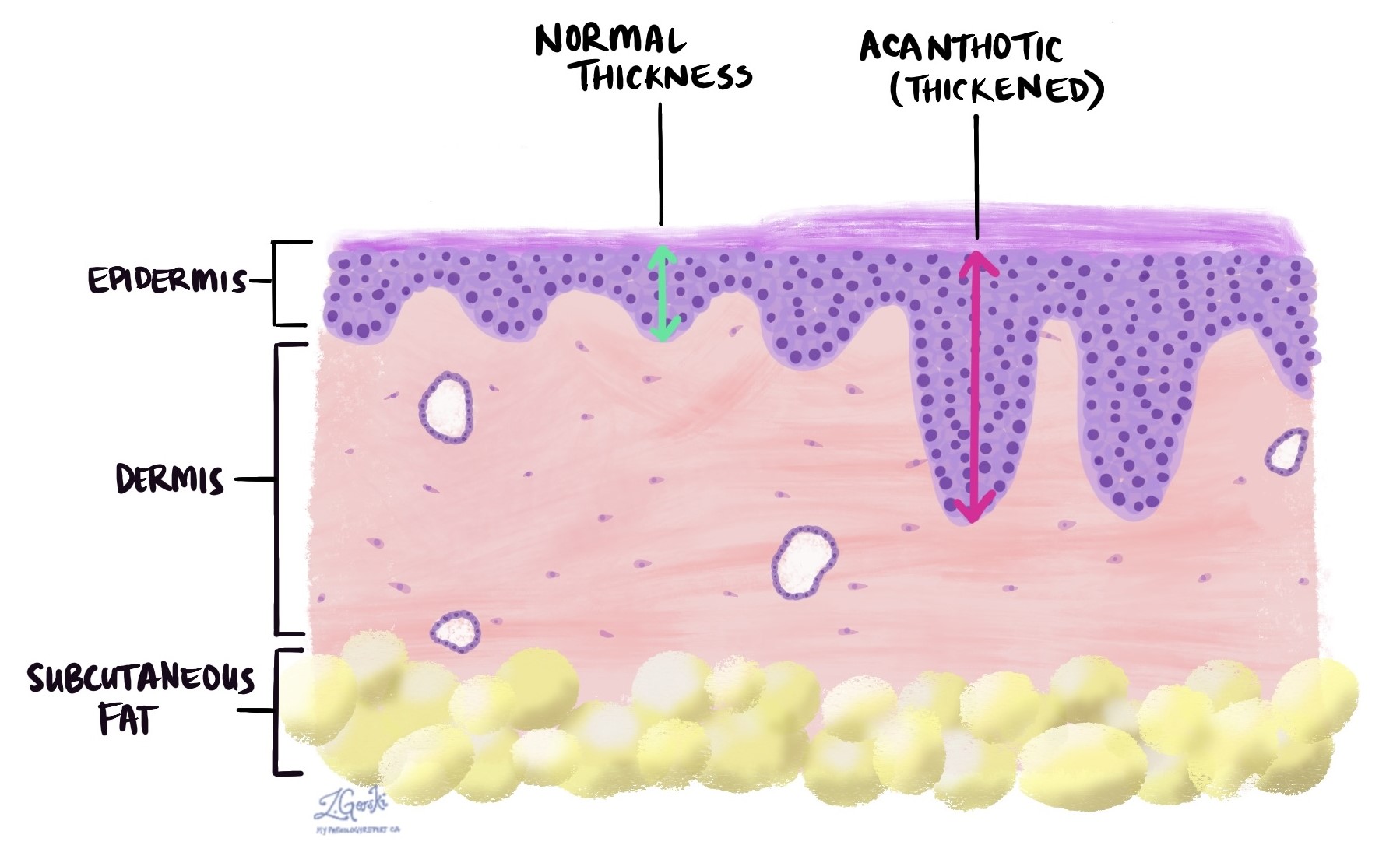
What does acanthosis look like under the microscope?
Under the microscope, acanthosis appears as a noticeable thickening of the squamous epithelium — the layer of flat cells that covers the surface of the tissue. The thickening is caused by extra cell layers, particularly in the middle portion of the epithelium. In benign acanthosis, the cells are usually normal in shape and well-organised, which helps the pathologist recognise that the change is reactive rather than cancerous.
Acanthosis is often accompanied by other microscopic changes. Keratosis — an increased buildup of the protective protein keratin on the surface — is a frequent companion finding. Signs of inflammation in the underlying tissue may also be present, reflecting whatever is driving the thickening.
Is acanthosis serious?
On its own, acanthosis is not serious and does not mean cancer is present. It is most often a straightforward protective reaction that requires no specific treatment beyond addressing the underlying cause — such as managing an inflammatory skin condition or reducing exposure to an irritant.
However, if acanthosis is found alongside other abnormal features — such as dysplasia (precancerous changes), an ulcer, or a mass — your doctor may recommend further tests or follow-up. The significance of acanthosis always depends on the full pathology report, the location of the tissue, and your clinical history.
Questions to ask your doctor
- What is causing the acanthosis in my report?
- Were there any other abnormal findings — such as dysplasia or inflammation — alongside the acanthosis?
- Do I need any follow-up tests or monitoring?
Related articles on MyPathologyReport.com
We are proud to partner with:
![]()