

An atypical mitotic figure is a cell caught in the act of dividing abnormally. Cells normally make copies of themselves through a carefully controlled process called mitosis, which evenly divides the cell’s genetic material (DNA) so that each new cell receives a complete and identical set. A dividing cell seen under the microscope is called a mitotic figure. When the division is disordered, so that the DNA is not being shared evenly, pathologists call it an atypical mitotic figure. This article explains what an atypical mitotic figure is, what it looks like under the microscope, and why it may appear in your pathology report.
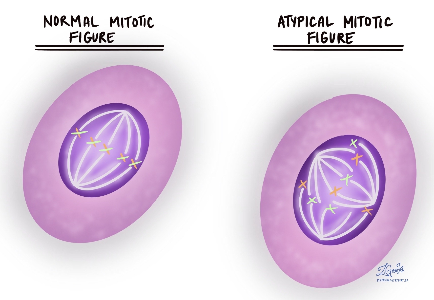
What does an atypical mitotic figure look like under the microscope?
During a normal cell division, the chromosomes (the structures that carry the DNA) line up neatly in the middle of the cell and are then pulled apart toward two opposite ends, like a tug-of-war with two evenly matched sides. This produces two new cells with matching sets of DNA. An atypical mitotic figure looks different because this orderly process has broken down. Instead of being pulled toward two ends, the chromosomes may be pulled toward three or more directions at once (described as tripolar or multipolar), arranged in a ring shape, or scattered and disorganized. The result is a division that cannot share the DNA evenly, so the new cells end up with too much, too little, or jumbled genetic material.
Why are atypical mitotic figures important?
Atypical mitotic figures matter because they indicate that cells are dividing in an uncontrolled, disorderly way, a feature of cancer. Uneven sharing of DNA during division leads to cells with an abnormal amount of genetic material, and this kind of genetic instability is one of the ways cancers grow and change over time. Because atypical mitotic figures are uncommon in normal tissue, their presence is a useful clue that helps a pathologist recognize and diagnose cancer. They also contribute to the tumor’s grade, a measure of how abnormal the cells look and how quickly they appear to be growing, which in turn helps the care team understand how the tumor is likely to behave.
Do atypical mitotic figures always mean cancer?
Not on their own. Atypical mitotic figures strongly point toward cancer, but their presence alone is not enough to make the diagnosis. Abnormal-looking divisions can occasionally appear in tissue that has been damaged by radiation therapy, chemotherapy, or certain infections, and rarely in some noncancerous conditions. For this reason, a pathologist always weighs this finding alongside the other features of the tissue and sometimes additional tests before reaching a final diagnosis.
Where are atypical mitotic figures found?
Atypical mitotic figures are most often seen in cancers, including common types such as breast cancer, lung cancer, colon cancer, and melanoma. They are usually absent from healthy tissue. Normal tissues that renew themselves quickly, such as the lining of the intestines or the bone marrow, do contain ordinary (normal) mitotic figures, but the abnormal, atypical forms are characteristic of disease.
What happens after an atypical mitotic figure is found?
When atypical mitotic figures are identified, the pathologist examines them alongside the other microscopic features of the tissue and may order additional tests, such as immunohistochemistry (IHC), to determine the type and origin of the abnormal cells. Immunohistochemistry uses markers that highlight specific proteins, helping confirm which types of cells are present. Together, these findings contribute to the final diagnosis and, when cancer is confirmed, to the tumor’s grade. The diagnosis, grade, type, and stage are what the care team uses to plan next steps and discuss treatment options, rather than the presence of atypical mitotic figures by itself.
Questions to ask your doctor
- What does the finding of atypical mitotic figures mean in my case?
- Were atypical mitotic figures part of how my diagnosis was made?
- Did this finding affect the grade of my tumor?
- Were any additional tests, such as immunohistochemistry, performed on my sample?
- Could this finding be related to previous radiation or chemotherapy rather than cancer?
- What do the diagnosis and grade tell us about how this condition is likely to behave?
- What are the next steps based on my overall pathology results?
We are proud to partner with:
![]()