

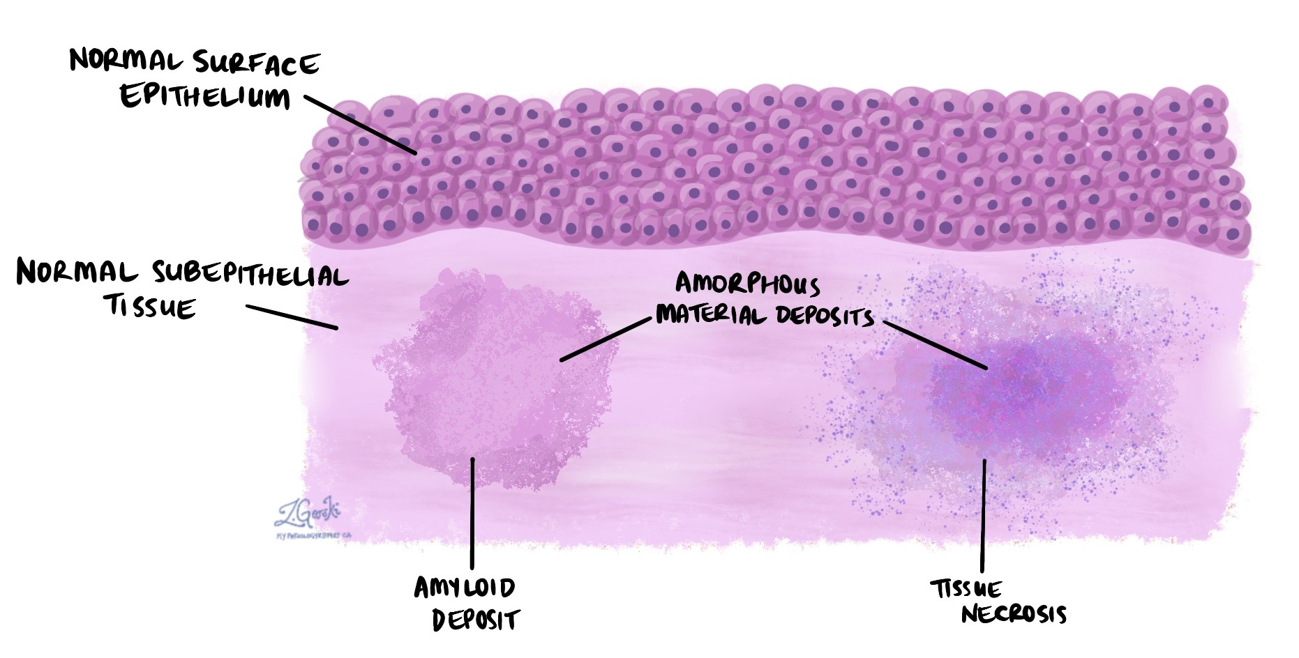
In pathology, the term amorphous is used to describe material that has no clear shape or structure when viewed under a microscope. Unlike normal tissues and cells, which have distinct forms and patterns, amorphous material appears shapeless and unorganized, often as a featureless background or a dense mass with no identifiable cell types.
This word is often used in pathology reports to describe cellular debris, protein deposits, or areas of tissue damage, and it can be an important clue to understanding what is happening in a tissue sample.
When do pathologists see amorphous material?
Amorphous material can be seen in a variety of medical conditions, including:
-
Necrosis – areas of tissue death, where the normal structure has broken down.
-
Amyloidosis – a condition where a protein called amyloid builds up in tissues as unstructured deposits.
-
Calcifications – sometimes calcium deposits may appear as amorphous debris.
-
Tumours – some cancers or benign tumours may contain amorphous material in the background, especially when cells break down or form unusual substances.
-
Inflammation or infection – damaged cells and inflammatory proteins can create an amorphous appearance in areas of chronic inflammation.
In each of these settings, the presence of amorphous material tells the pathologist that normal tissue architecture has been disrupted.
Why is amorphous material important?
Even though amorphous material has no specific shape or pattern, it can still offer important diagnostic information. For example:
-
In amyloidosis, identifying amorphous protein deposits helps confirm the diagnosis.
-
In necrosis, amorphous areas can help identify where tissue has died, which may be related to infection, trauma, or lack of blood supply.
-
In tumours, amorphous material may reflect areas of cell breakdown or provide insight into the tumour’s composition and behavior.
In some cases, the amorphous material is the main clue that helps the pathologist determine the type or severity of a disease.
How do pathologists study amorphous material?
Because amorphous material lacks obvious structure, pathologists often use special stains to learn more about what it’s made of:
-
Congo red stain – used to detect amyloid deposits, which show a pink color under regular light and an apple-green glow under polarized light.
-
PAS (Periodic acid–Schiff) – highlights certain carbohydrates and protein deposits.
-
Calcium stains – such as von Kossa or Alizarin Red, used to detect mineral deposits.
-
Immunohistochemistry or molecular testing – sometimes used to determine whether the amorphous material contains specific proteins or tumour markers.
These tools help pathologists identify what the amorphous material is and whether it is related to a specific disease.
Questions to ask your doctor
-
What does the term “amorphous” mean in my pathology report?
-
Was the amorphous material linked to a particular diagnosis?
-
Do I need further testing or follow-up?
-
Could the presence of this material affect my treatment plan?
We are proud to partner with:
![]()