by Jason Wasserman MD PhD FRCPC
May 16, 2024
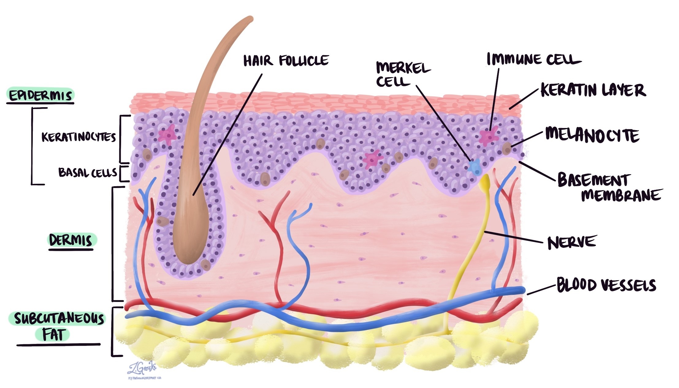
A blue nevus is a type of benign (noncancerous) growth made up of specialized cells called melanocytes. It is characterized by its distinctive blue or bluish-gray color, which is caused by the presence of melanin deep within the dermis, a phenomenon known as the Tyndall effect.

What causes a blue nevus?
Blue nevus is generally considered a congenital condition that occurs from birth or develops in early childhood. However, some cases may appear later in life. The nevus is formed by the accumulation of melanocytes (the melanin-producing cells) in the deeper layers of the skin, which is somewhat atypical since melanocytes are usually located in the upper layers of the skin.
What does a blue nevus look like?
Visually, a blue nevus is typically small, round, or oval and has a smooth surface. Its distinctive blue color is due to the melanin being deeper in the dermis rather than in the epidermis, causing light scattering and absorption, which gives the nevus its blue appearance.
Microscopic features
Under the microscope, a blue nevus is characterized by:
- Spindle-shaped melanocytes: These cells are usually densely packed and located deep within the dermis.
- Melanin pigmentation: The melanocytes in a blue nevus contain a significant amount of melanin, contributing to the lesion’s color.
- Lack of junctional activity: Unlike some other types of nevi, blue nevi typically do not show an active junctional component, where melanocytes cluster at the dermo-epidermal junction.
Does a blue nevus need to be removed?
Generally, a blue nevus does not require removal unless it changes in size, shape, or color or becomes symptomatic (itching, bleeding, etc.), which could be signs of malignancy. Regular monitoring is usually sufficient. In cases where there is uncertainty about the diagnosis or concerns about cosmetic appearance, a dermatologist might recommend removal to ensure there is no risk of melanoma. The decision to remove a blue nevus should be based on clinical judgment and patient preference, especially if the nevus poses a cosmetic concern or there is anxiety about its nature.
About this article
Doctors wrote this article to help you read and understand your pathology report. Contact us if you have any questions about this article or your pathology report. Read this article for a more general introduction to the parts of a typical pathology report.


