

by Robyn Ndikumana MD BScN and Allison Osmond, MD FRCPC
November 14, 2024
A congenital nevus is a common, non-cancerous skin tumour made up of melanocytes, cells responsible for producing melanin, the pigment that gives skin its colour. Congenital nevi (the plural of nevus) are usually present at birth or develop within the first year of life, which is why they are called “congenital.” Another name for this type of growth is a mole. The term “mole” is often used to describe any skin growth of melanocytes.
Congenital nevi are more common in people with light-coloured skin, and they can be found anywhere on the body, though they most often appear on the trunk and limbs.
What causes a congenital nevus?
A congenital nevus forms when melanocytes overgrow during fetal development. This overgrowth happens because of changes in specific genes that control cell growth, causing clusters of melanocytes to develop in one area of the skin. These genetic changes occur randomly and are not inherited from a parent or caused by anything the mother did during pregnancy.
Can a congenital nevus turn into melanoma over time?
It is very rare for a congenital nevus to turn into melanoma, a type of skin cancer, over time. The risk is highest for very large congenital nevi (over 20 cm), but even in these cases, the chance of developing melanoma is low. Routine monitoring and self-examination can help detect any changes that could be signs of melanoma.
How common are congenital nevi?
Congenital nevi are relatively common, with about 1% of newborns having small congenital nevi. Larger nevi, those measuring over 1.5 cm, are much less common. Giant congenital nevi, defined as those larger than 20 cm, are rare.
What does a congenital nevus look like?
Congenital nevi can vary significantly in appearance. They range from very small to very large, with giant congenital nevi measuring over 20 cm. Most congenital nevi are irregular in shape, and their colour can range from light brown to black. It is not uncommon for hair to grow out of a congenital nevus, which is a normal characteristic and does not indicate any health issues.
How is this diagnosis made?
A congenital nevus is often diagnosed after the growth has been surgically removed and sent to a pathologist for examination. Sometimes, a doctor can recognize this growth by its appearance. Still, a biopsy or removal is sometimes needed to confirm the diagnosis and rule out other skin conditions.
Microscopic features
When examined under a microscope, a congenital nevus has distinct features that help pathologists identify it:
- Melanocytes: The tumour primarily comprises melanocytes, the cells that produce melanin pigment. These cells often contain dark melanin pigment, giving the tumour its colour.
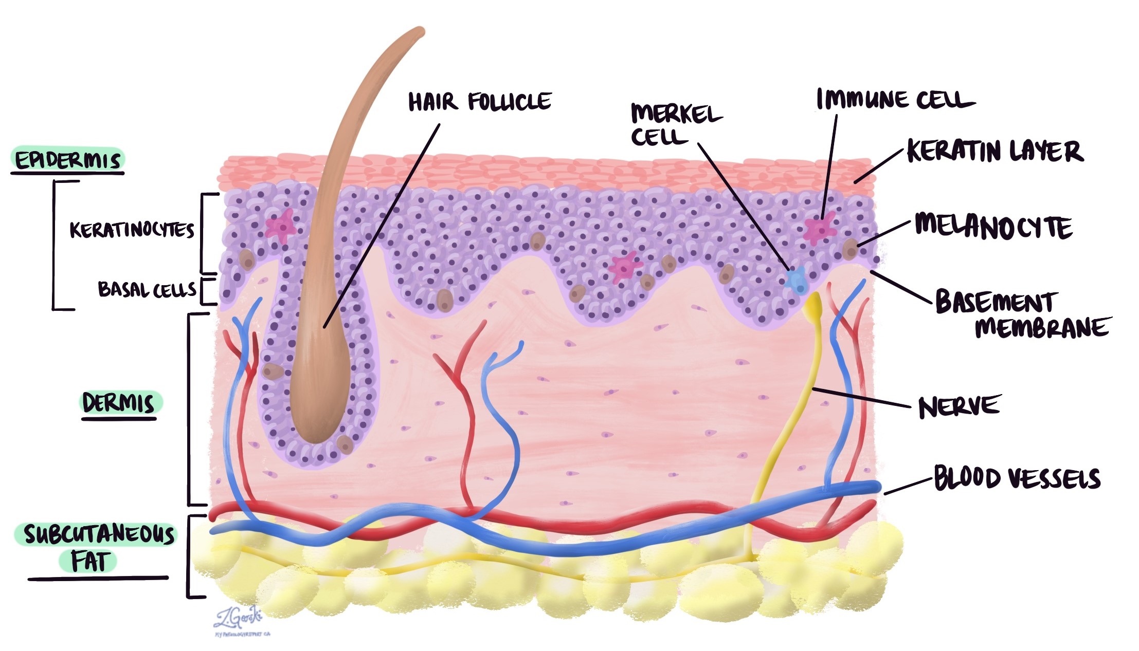
- Location in the skin: The melanocytes in a congenital nevus grow primarily in the dermis, which is the layer of skin just below the surface.
- Growth around normal structures: As the melanocytes in a congenital nevus grow, they may surround normal structures in the skin, such as hair follicles, sebaceous (oil) glands, nerves, and blood vessels. This growth pattern is typical for this type of growth and helps distinguish them from other moles.
- Involvement of deeper tissues: In some cases, melanocytes can grow into the layer of fat beneath the dermis, known as the subcutaneous tissue. This feature is more common in larger nevi.

We are proud to partner with:
![]()