by Iris Teo, MD FRCPC
November 25, 2024
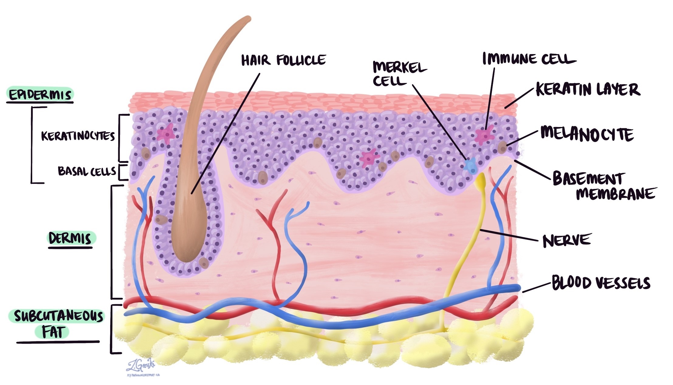
An epidermoid cyst is a non-cancerous growth that develops below the skin’s surface. It is a round, hollow structure lined by the same squamous cells found in a layer of the skin called the epidermis. Epidermoid cysts are sometimes referred to as ‘epidermal cysts’, ‘infundibular cysts’, or ‘epidermal inclusion cysts.’

Where are epidermoid cysts typically found?
Epidermoid cysts are frequently found on the face, neck, and torso.
What causes an epidermoid cyst?
No one knows why epidermoid cysts occur. On some areas of the body, like the palms and soles, they may be caused by trauma that pushes some of the epidermis into the dermis. At other body sites, they are thought to arise from inflammation of the hair follicle.
How is this diagnosis made?
The diagnosis is usually made after the cyst is surgically removed and sent to a pathologist for examination under the microscope.
What does an epidermoid cyst look like under the microscope?
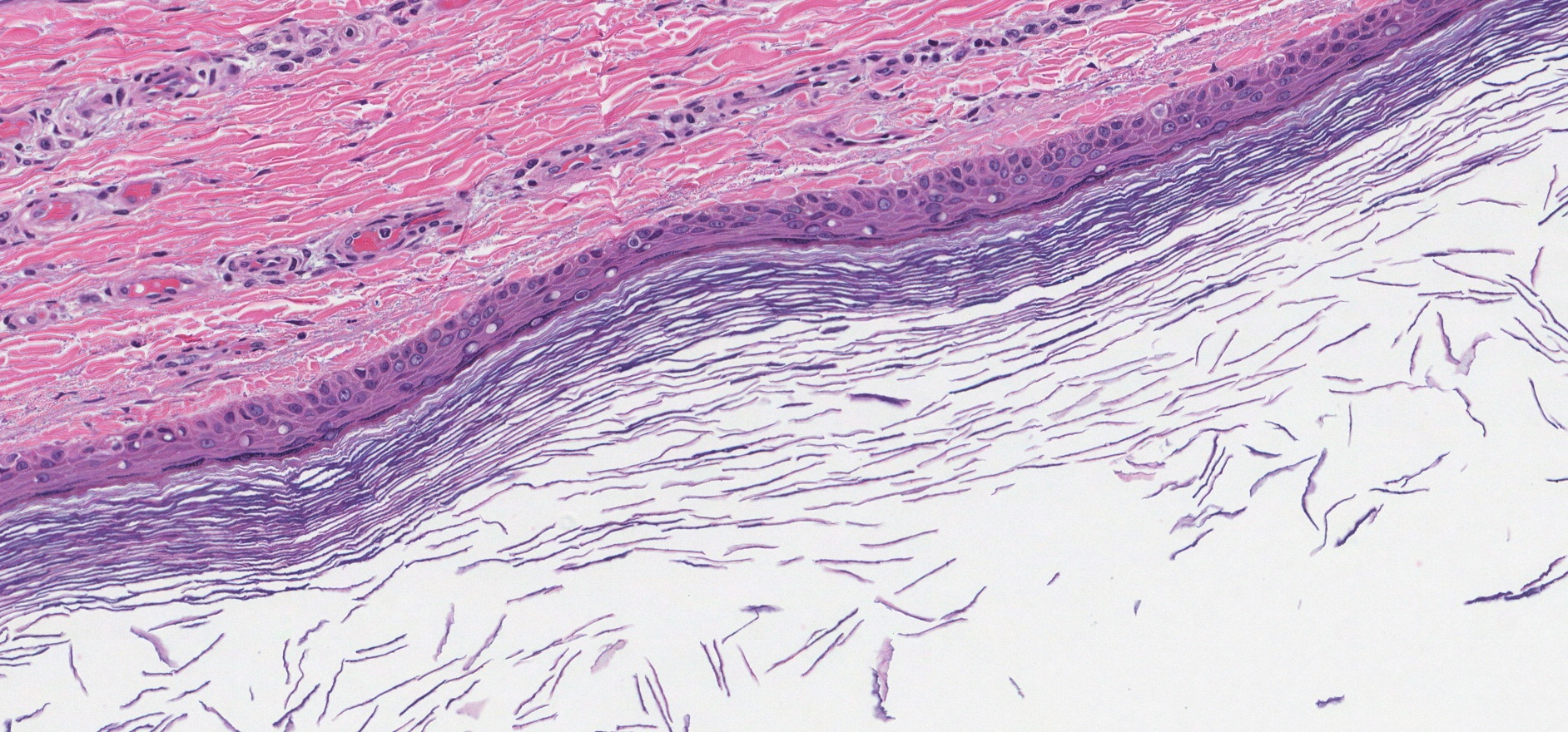
When examined under the microscope, an epidermoid cyst is a round structure lined by squamous cells. The squamous cells show a maturation similar to that seen on the skin surface. The inside of the cyst may be filled with keratin debris, which looks similar to the keratin found on the surface of the skin.
Epidermoid cysts may break open or rupture. This can lead to pain and inflammation as keratin inside the cyst spills into the surrounding tissue. When examined under the microscope, your pathologist may see specialized immune cells called multi-nucleated giant cells and other chronic inflammatory cells in the dermis around the ruptured cyst.


