

by Jason Wasserman MD PhD FRCPC and Zuzanna Gorski MD
August 13, 2026
Fibrocystic change is a noncancerous (benign) condition of the breast made up of several related changes that often occur together, including cysts, fibrosis, apocrine metaplasia, and adenosis. It is very common, affecting up to 60% of women of reproductive age, and it can involve one or both breasts.
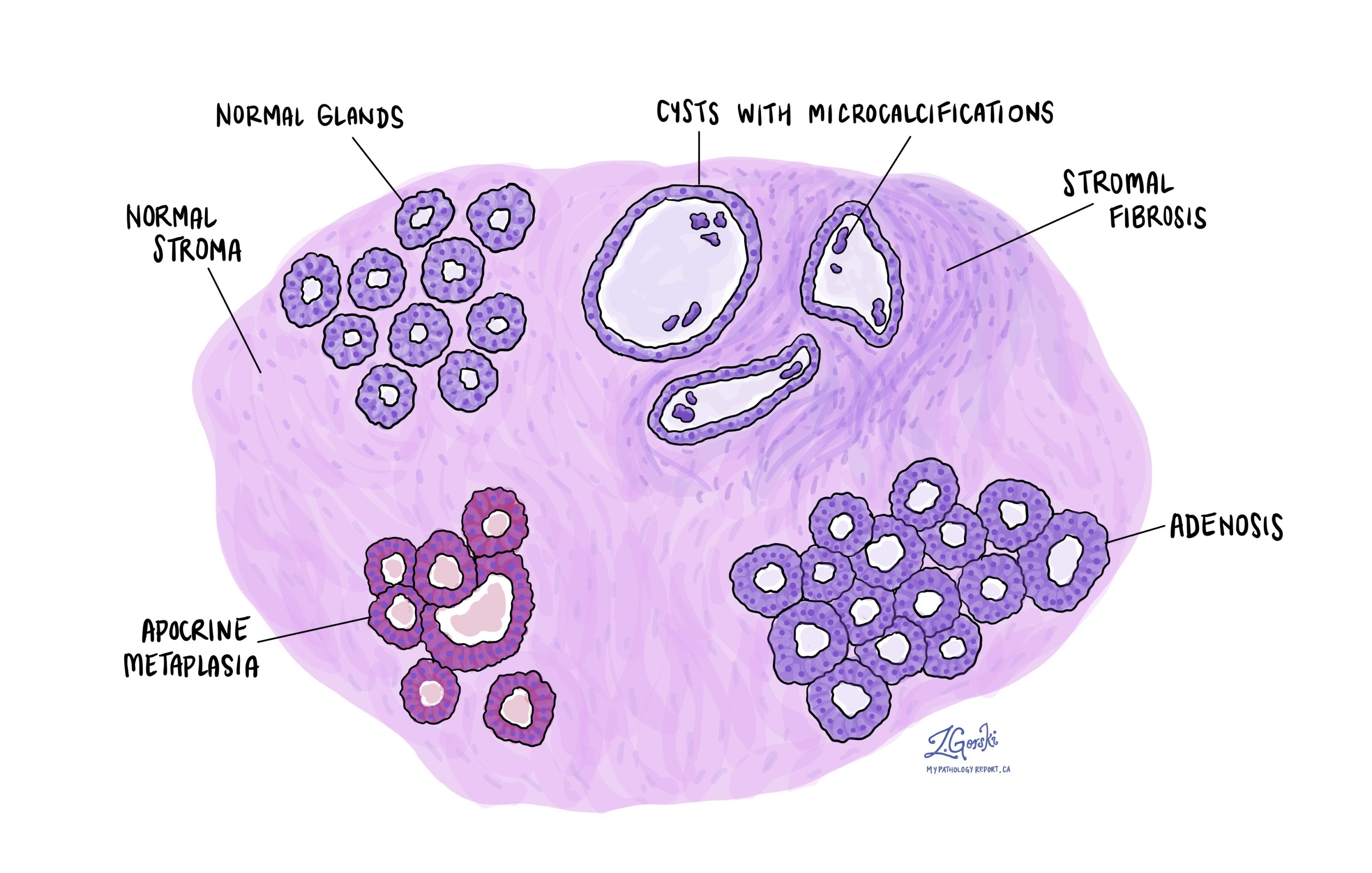
Fibrocystic change is not a disease, and the term “fibrocystic change” is preferred over the older name “fibrocystic disease” for that reason. It is best understood as a description of the normal breast responding to hormones over many years. This article will help you understand the findings in your pathology report, what each term means, and why it matters for your care.
What are the symptoms of fibrocystic change?
Fibrocystic change may cause lumpiness, tenderness, or discomfort in one or both breasts, and these symptoms are often most noticeable in the days before menstruation. Some people feel a distinct lump, which is usually a large cyst. Many people have no symptoms at all, and the changes are found only when breast tissue is examined for another reason.
What causes fibrocystic change?
Fibrocystic change is thought to develop in response to hormonal stimulation, particularly from the hormones estrogen and progesterone, which normally rise and fall during the menstrual cycle. These hormones cause the breast ducts and glands to enlarge and then shrink, and over many cycles this leads to the development of cysts and fibrous (scar-like) tissue. Because the changes are hormone-driven, symptoms often improve after menopause.
How is the diagnosis made?
The diagnosis of fibrocystic change is made after breast tissue is examined under the microscope by a pathologist. The sample is typically obtained from a core needle biopsy performed because an abnormal area, such as a density or a calcification, was seen on a mammogram or ultrasound. Fibrocystic change is also often found incidentally in tissue removed for another reason.
Under the microscope, fibrocystic change is made up of several features that may appear together or separately. Your report may mention any combination of them.
- Cysts — A cyst is a round or oval sac filled with fluid. In fibrocystic change, normally small breast glands enlarge and fill with fluid. Large cysts may be felt as a lump, and calcium deposits can form within them, appearing as bright white spots on a mammogram.
- Stromal fibrosis — The stroma is the supportive tissue around the glands and ducts. When a cyst breaks open, fluid leaks into the stroma, causing inflammation and activating fibroblasts, the cells that produce connective tissue. The result is dense, scar-like tissue, a process called fibrosis, which can make parts of the breast feel firm or rope-like.
- Apocrine metaplasia — Metaplasia means one type of mature cell changes into another. In apocrine metaplasia, the normal duct lining cells change into apocrine cells, which are larger, with a round nucleus and pink cytoplasm. This is a common and noncancerous part of fibrocystic change.
- Adenosis — Adenosis means there are more glands than usual in one area of the breast, and the glands may be larger than normal. It often occurs together with columnar cell change and columnar cell hyperplasia, which are mild, noncancerous alterations in the cells lining the ducts. When the extra glands are surrounded by scar-like tissue that distorts them, the change is called sclerosing adenosis.
Does fibrocystic change increase the risk of breast cancer?
For most people, fibrocystic change does not increase the risk of developing breast cancer. Fibrocystic change is an umbrella term, however, and the answer depends on which specific changes the pathologist found in your sample. Pathologists group them as follows:
- Non-proliferative changes — Cysts, fibrosis, and apocrine metaplasia make up the large majority of fibrocystic change. These are not associated with an increased risk of breast cancer.
- Proliferative changes without atypia — Changes involving an increase in the number of normal-looking cells, such as usual ductal hyperplasia and sclerosing adenosis, are associated with a small increase in risk. The overall risk remains low.
- Proliferative changes with atypia — When atypical ductal hyperplasia or atypical lobular hyperplasia is found, the increase in risk is greater, and these findings are managed with closer follow-up. They are separate diagnoses rather than part of fibrocystic change itself.
Because some areas of fibrocystic change can feel like a lump or produce calcifications on imaging, a biopsy is often performed to confirm that the changes are benign. Once that is confirmed under the microscope, no further treatment is usually needed.
What happens after this diagnosis?
The outlook for fibrocystic change is excellent. It is a benign condition, it does not turn into cancer, and many people find that symptoms improve after menopause when hormone levels fall. Treatment is usually not necessary. When tenderness or lumpiness is bothersome, supportive measures such as wearing a well-fitting supportive bra, reducing caffeine, or using mild pain relief may help. A large or uncomfortable cyst can sometimes be drained with a needle. Your doctor may recommend continuing with routine breast imaging so that any new change can be assessed, and will discuss follow-up if the report also mentioned a proliferative or atypical change.
Questions to ask your doctor
- Do my biopsy results show any findings besides fibrocystic change?
- Were any proliferative or atypical changes, such as usual ductal hyperplasia, sclerosing adenosis, or atypical ductal hyperplasia, found?
- Does my result change my future risk of breast cancer?
- Are there calcifications or cysts that need follow-up?
- Did my biopsy result match my imaging findings?
- Do I need another imaging test or biopsy in the future?
- Is there anything I can do to help with breast tenderness or lumpiness?
- What symptoms should prompt me to come back for re-evaluation?
Related articles on MyPathologyReport.com
- Sclerosing Adenosis of the Breast: Understanding Your Pathology Report
- Usual Ductal Hyperplasia (UDH): Understanding Your Pathology Report
- Columnar Cell Change of the Breast: Understanding Your Pathology Report
- Fibroadenoma of the Breast: Understanding Your Pathology Report
- Atypical Ductal Hyperplasia (ADH): Understanding Your Pathology Report
- Understanding Your Breast Biopsy Pathology Report
We are proud to partner with:


🔍 Search MyPathologyReport
Type what you see on your report — for example a diagnosis or test name