

by Robyn Ndikumana MD BScN and Allison Osmond, MD FRCPC
November 25, 2024
A hemangioma is a non-cancerous tumour made up of an abnormal collection of blood vessels. These tumours often appear red to blue and typically have a well-defined border separating them from the surrounding normal tissue. Hemangiomas can occur anywhere on the body but are most commonly found on the skin, head, neck, and liver. The size of a hemangioma can range widely, from very small (1–2 millimetres) to very large (over 20 centimetres).
At what age does a hemangioma develop?
Hemangiomas can develop at any age. Tumours present at birth are called congenital hemangiomas. In contrast, infantile hemangiomas appear within the first few days or weeks after birth. Some other types of hemangiomas may develop later in life.
What are the symptoms of a hemangioma?
The symptoms of a hemangioma depend on its location, size, and type. Common symptoms include:
- Visible red or blue lump: Most hemangiomas on the skin are easily noticeable and may grow in size before stabilizing or regressing.
- Pain or tenderness: Hemangiomas near nerves or sensitive tissues may cause discomfort or pain.
- Swelling or pressure effects: Large hemangiomas in certain areas, such as the liver or brain, may press on nearby structures, leading to functional symptoms.
- Bleeding: Superficial hemangiomas can sometimes break open, especially if injured, leading to bleeding.
- Cosmetic concerns: Depending on their location, hemangiomas can sometimes cause cosmetic issues that may affect a person’s confidence or self-esteem.
Some hemangiomas may not cause symptoms and are only discovered during imaging studies or other medical evaluations.
Can a hemangioma become cancerous?
Hemangiomas are benign (non-cancerous) tumours that do not transform into cancer. However, very rarely, a lesion that resembles a hemangioma may be a different condition, such as angiosarcoma (a rare cancer of the blood vessels). For this reason, it is important to have any unusual or persistent growth evaluated by a doctor to confirm the diagnosis and rule out other possibilities.
What types of blood vessels may be found in a hemangioma?
Hemangiomas are made up of abnormal blood vessels, which can include capillaries, veins, and arteries. The type of blood vessels present depends on the specific kind of hemangioma:
- Capillaries: These are the smallest blood vessels, responsible for connecting arteries to veins and enabling the exchange of oxygen and nutrients with surrounding tissues. Capillary hemangiomas are composed primarily of tightly packed capillaries, which give the tumour its bright red colour and characteristic appearance.
- Veins: These larger vessels carry blood back to the heart. Veins found in hemangiomas typically have thinner walls and may form broad, irregular spaces filled with blood. Cavernous hemangiomas often contain prominent venous structures, contributing to their deeper and more expansive growth.
- Arteries: These blood vessels carry oxygen-rich blood away from the heart. While less common, hemangiomas can sometimes include small arteries or arterioles. These vessels may be present in certain types, such as epithelioid hemangiomas, and can contribute to increased blood flow within the tumour.
The specific combination and structure of these blood vessels help pathologists determine the type of hemangioma and guide treatment decisions.
Types of hemangiomas
Hemangiomas are classified based on when they develop, their location on the body, and the types of blood vessels found within the tumour. All hemangiomas are non-cancerous, though their behaviour can vary. Some may disappear on their own over time, while others might require surgical removal. Common types include:
- Capillary hemangioma: Often called “strawberry hemangioma” due to its red colour, this type typically forms in the skin and often regresses over time.
- Cavernous hemangioma: Found in organs like the liver, brain, or eye, these hemangiomas do not regress and, depending on their size and location, may require treatment.
- Congenital hemangioma: Present at birth, these hemangiomas may either regress naturally or persist, depending on their type.
- Epithelioid hemangioma: This tumour, which typically occurs in the head and neck area, may arise after an injury to the skin. Under the microscope, it often shows a significant number of immune cells called eosinophils. It is also known as angiolymphoid hyperplasia with eosinophils.
- Targetoid hemangioma: Also known as a “hobnail hemangioma”, it resembles a ‘target’ with a red or blue centre surrounded by a pale or dark rim.
- Infantile hemangioma: Developing in infancy, these hemangiomas are common and often undergo regression without treatment.
- Lobular capillary hemangioma: These fast-growing tumours can mimic cancer. They are often linked to injuries or pregnancy. Previously called “pyogenic granulomas,” this term is misleading as these tumours are neither ‘pyogenic’ (pus-forming) nor ‘granulomatous.’
- Verrucous hemangioma: Usually appearing soon after birth, these hemangiomas resemble a bump or wart on the skin.
- Infiltrating hemangioma: Unlike other types, infiltrating hemangiomas lack a clear border, making complete surgical removal challenging.
What types of blood vessels may be found in a hemangioma?
The types of blood vessels found in a hemangioma depend on the specific type of tumour. Common types of blood vessels include:
- Capillaries: These are small, thin-walled vessels often found in capillary hemangiomas. They are arranged closely together, giving the tumour its characteristic red colour.
- Larger blood vessels: Cavernous hemangiomas contain larger, thicker-walled vessels, which may be irregularly shaped. These vessels form deeper and broader spaces within the tumour.
- Atypical vessels: Some hemangiomas, such as epithelioid or targetoid hemangiomas, may show blood vessels with unusual shapes or patterns, which can help their identification under a microscope.
The type of blood vessel present helps pathologists classify the hemangioma and distinguish it from other vascular conditions.
How is the diagnosis of hemangioma made?
The diagnosis of hemangioma is typically made by examining a small sample of the tumour removed during a biopsy or after the complete tumour has been surgically excised. The tissue is sent to a pathologist, who examines it under a microscope to confirm the diagnosis.
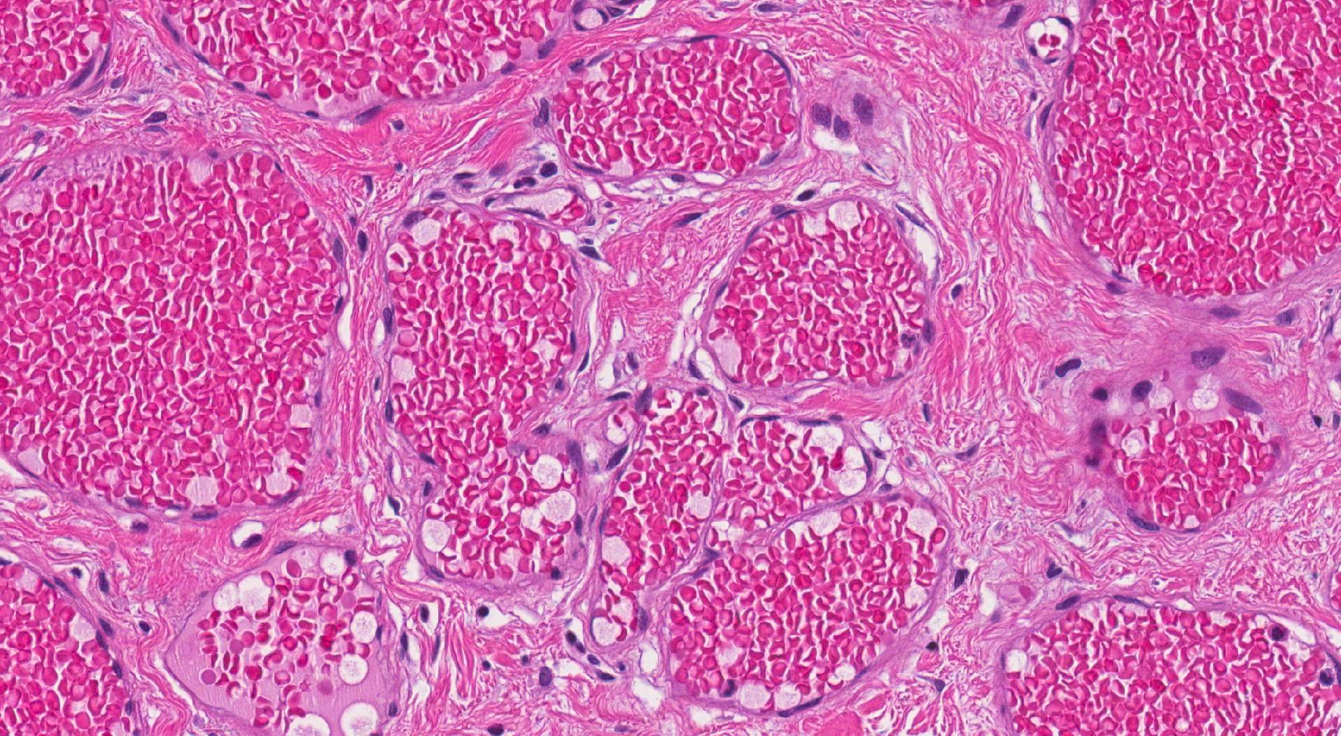
What are the microscopic features of a hemangioma?
When examined under a microscope, hemangiomas are composed of abnormal blood vessels that may vary in size and shape depending on the type of hemangioma. These vessels are lined by endothelial cells, which form the inner lining of blood vessels and are supported by surrounding connective tissue.
What does tumour regression mean?
Tumour regression refers to a natural decrease in the size of a tumour, often to the point of complete disappearance. Regression is commonly seen in infantile hemangiomas, which may shrink or vanish as a child grows older. Tumours that do not regress may require treatment, which could include surgical removal or medications designed to shrink the tumour.
Does a hemangioma need to be removed?
Not all hemangiomas require treatment. Whether a hemangioma needs to be removed depends on its size, location, symptoms, and potential for complications.
- No treatment needed: Many infantile hemangiomas regress over time and do not require medical or surgical intervention.
- Medication: For hemangiomas that are causing symptoms, medications such as beta-blockers (e.g., propranolol) can help shrink the tumour.
- Surgical removal: Surgery may be recommended for hemangiomas that do not regress, interfere with function, cause pain, or pose cosmetic concerns. In cases where removal is challenging, other therapies, such as laser treatment, may also be considered.
Your doctor will assess the hemangioma and discuss the best management approach based on your circumstances.
We are proud to partner with:
![]()