

by Jason Wasserman MD PhD FRCPC
July 26, 2025
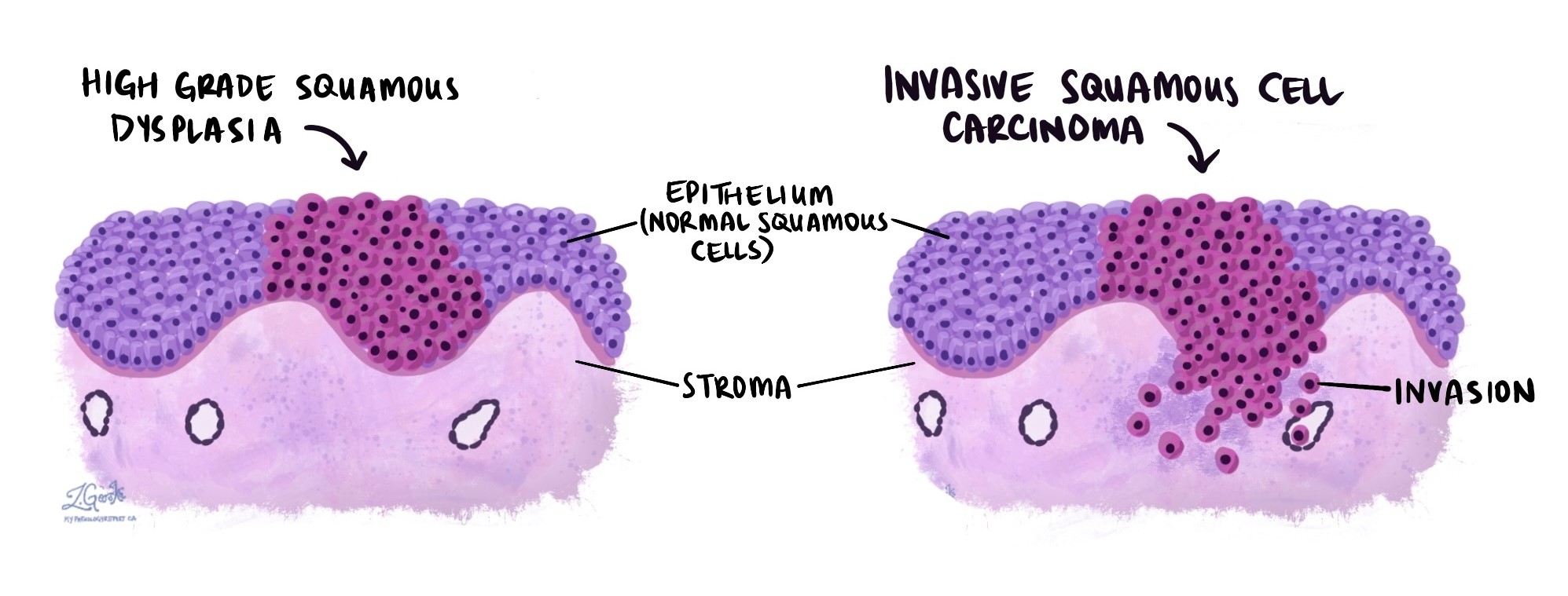
High grade squamous dysplasia is a precancerous condition that affects the squamous cells lining the inside of the esophagus (the tube that carries food from your mouth to your stomach). These squamous cells typically form a smooth, protective layer known as the squamous epithelium.
In high grade squamous dysplasia, the cells look significantly abnormal when examined under the microscope. These changes suggest the cells are growing in an uncontrolled way, which puts them at high risk of turning into a type of esophageal cancer called squamous cell carcinoma if left untreated.
What causes high grade squamous dysplasia in the esophagus?
High grade squamous dysplasia usually develops as a result of long-term damage or irritation to the squamous epithelium. Over time, repeated injury can cause the cells to become abnormal.
Common causes include:
-
Smoking – Tobacco use is one of the strongest risk factors for squamous dysplasia and squamous cell carcinoma.
-
Alcohol use – Drinking large amounts of alcohol can irritate the esophagus and increase the risk.
-
Chronic acid reflux – Gastroesophageal reflux disease (GERD) allows stomach acid to flow back into the esophagus, which can lead to inflammation and damage.
-
Chronic irritation – Repeated exposure to very hot liquids, spicy foods, or other chemical or physical irritants can injure the squamous cells.
-
Infections – In some parts of the world, viral infections like HPV (human papillomavirus) have been linked to squamous dysplasia in the esophagus.
Reducing these risk factors may lower the chance of progression from dysplasia to cancer.
What are the symptoms?
In most cases, high grade squamous dysplasia does not cause any noticeable symptoms. Because of this, it is often discovered by chance during a procedure such as an endoscopy performed for another reason.
If symptoms are present, they may include:
-
Difficulty swallowing (also called dysphagia).
-
Pain when swallowing.
-
Persistent heartburn or acid reflux.
-
A feeling that food is stuck in the throat or chest.
These symptoms are not specific to dysplasia and can occur with other conditions of the esophagus, including inflammation or cancer.
How is this diagnosis made?
High grade squamous dysplasia is diagnosed after your doctor performs an upper endoscopy, a procedure that allows them to look inside your esophagus using a thin, flexible tube with a camera on the end.
If an abnormal area is seen, a biopsy will be taken. A biopsy is a small tissue sample removed from the surface of the esophagus.
A pathologist examines the sample under a microscope to look for changes in the squamous cells. If the changes are severe enough, the diagnosis of high grade dysplasia is made.
What does high grade squamous dysplasia look like under the microscope?
When viewed under the microscope, high grade squamous dysplasia shows significant changes in the way the squamous cells look and how they are organized.
Cytological atypia (abnormal cells):
-
Large, darkly stained, irregular nuclei (the part of the cell that contains DNA).
-
Loss of normal cell shape and structure.
-
Crowding or overlapping of cells.
-
Increased number of mitoses (cells that are dividing).
Architectural atypia (abnormal tissue organization):
-
Disruption in the normal maturation of cells as they move from the bottom to the top of the squamous epithelium.
-
Abnormal thickening of the tissue.
High grade squamous dysplasia is diagnosed when these abnormalities involve more than half of the thickness of the squamous epithelium, or if the cell changes are particularly severe, even if only part of the epithelium is affected.
What is the risk of developing esophageal cancer?
High grade squamous dysplasia is considered the final stage before cancer (specifically, squamous cell carcinoma) develops. If not treated, the abnormal cells have a high risk of progressing to cancer over time.
Because of this risk, this condition is taken very seriously, even though it is not cancer yet.
Factors that may influence the risk of cancer progression include:
-
Smoking or continued alcohol use.
-
A history of chronic acid reflux or inflammation.
-
Family history of esophageal cancer.
-
How widespread or severe the dysplasia is.
How is high grade squamous dysplasia treated?
The goal of treatment is to remove or destroy the abnormal cells before they turn into cancer. Treatment options may include:
-
Endoscopic mucosal resection (EMR) – The abnormal area is removed during an endoscopy using special tools.
-
Radiofrequency ablation (RFA) – Heat energy is used to destroy the abnormal tissue.
-
Cryotherapy – Freezing the tissue to kill abnormal cells.
-
Esophagectomy – In rare or extensive cases, surgery to remove part or all of the esophagus may be considered.
Your doctor will help you decide on the best treatment based on the location and extent of the dysplasia, your overall health, and your preferences.
What happens after treatment?
After treatment, it is very important to have regular follow-up with your doctor, which may include:
-
Repeat endoscopies.
-
Additional biopsies.
-
Monitoring for recurrence or progression.
Even after successful treatment, there is still a small chance that new areas of dysplasia or cancer could develop.
Questions to ask your doctor
- What caused this condition in my case?
-
What are my treatment options?
-
Will I need repeated endoscopies or biopsies?
-
What is my risk of developing esophageal cancer?
-
What lifestyle changes can I make to reduce my risk?
We are proud to partner with:
![]()