

by Jason Wasserman MD PhD FRCPC
August 12, 2026
An intraductal papilloma is a noncancerous (benign) tumor that grows inside one of the small milk ducts of the breast. The word intraductal means “inside the duct,” and papilloma refers to the small finger-like projections, called papillae, that form within the duct. Intraductal papillomas are among the most common benign breast tumors and cannot spread to other parts of the body.
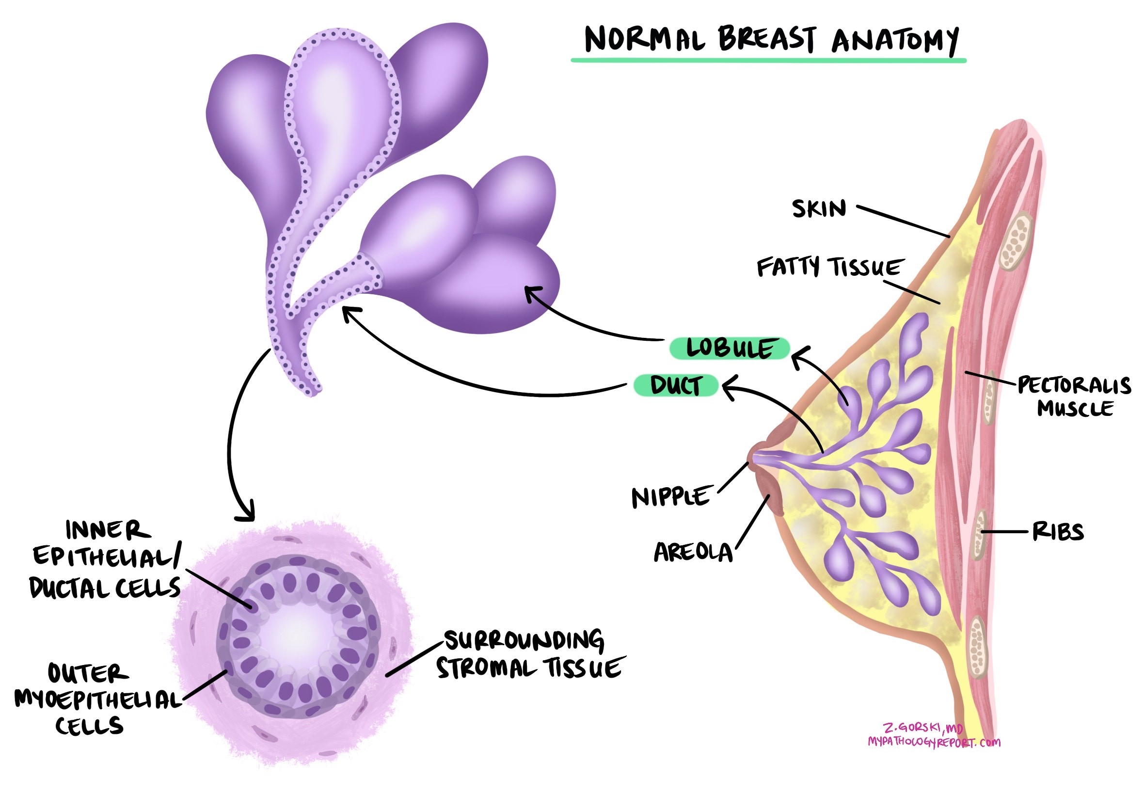
Where the papilloma grows affects how it behaves. A papilloma in the central part of the breast, just beneath the nipple, is usually solitary (a single tumor) and tends to be larger. Papillomas that form in the outer (peripheral) parts of the breast are often smaller, may be multiple, and are more likely to be linked with atypical changes. This article will help you understand the findings in your pathology report, what each term means, and why it matters for your care.
What are the symptoms of intraductal papilloma?
Many intraductal papillomas cause no symptoms and are found by chance on a mammogram or ultrasound. When symptoms do occur, the most common is nipple discharge, which may be clear or bloody and is especially common when the papilloma sits under or behind the nipple. Some people notice a small lump or thickened area beneath the nipple, and occasionally there is tenderness. Papillomas located deeper in the breast rarely cause discharge and are usually too small to feel.
What causes an intraductal papilloma?
The exact cause of an intraductal papilloma is not known. Most cases are sporadic, meaning they occur by chance and are not inherited. Hormonal influences, particularly from estrogen, are thought to play a role, since these tumors appear most often in women between the ages of 35 and 55.
How is the diagnosis made?
An intraductal papilloma is diagnosed after breast tissue is examined under the microscope by a pathologist. The tissue is usually obtained from a core needle biopsy or after surgical removal of a lump or an abnormal area seen on imaging. Under the microscope, an intraductal papilloma is made up of finger-like papillary structures that project into a duct. These structures are built around thin cores of connective tissue and small blood vessels (called fibrovascular cores) and are lined by two layers of cells: an inner layer of ductal epithelial cells and an outer layer of myoepithelial cells (specialized cells that form a supporting layer around the ducts).
The presence of a continuous layer of myoepithelial cells is one of the key features that separates a benign papilloma from a papillary carcinoma (a cancerous papillary tumor), which lacks this layer. To make this distinction clear, the pathologist often performs immunohistochemistry (IHC), a test that uses antibodies to highlight the myoepithelial cells and confirm that the lesion is benign.
Other findings within an intraductal papilloma
A pathology report for an intraductal papilloma may describe additional changes within the tumor. Some are benign and do not increase the risk of cancer, while others are atypical and do.
- Benign changes — Usual ductal hyperplasia (UDH), an increase in normal-looking duct-lining cells, and apocrine change, in which the duct cells take on features of sweat gland cells, are common and part of the normal spectrum of benign breast changes. They do not increase the risk of cancer.
- Atypical changes — In some papillomas, particularly peripheral ones, atypical ductal hyperplasia (ADH) or ductal carcinoma in situ (DCIS) can develop within the tumor. This means some of the duct cells have begun to grow abnormally, though they have not invaded the surrounding tissue. When ADH or DCIS is present, the papilloma is usually removed completely.
What happens after this diagnosis?
Because an intraductal papilloma is benign, the outlook is excellent, and once it is completely removed it rarely comes back. What happens next depends mainly on whether atypical changes are present and on whether the biopsy result matches the imaging. The pathology findings guide which options your team discusses, rather than dictating a single path.
- Surgical removal — Removing the papilloma is often recommended, especially when a core needle biopsy shows atypical ductal hyperplasia or DCIS, when the biopsy result does not match the imaging, or when the papilloma causes symptoms such as bloody nipple discharge. This is partly because a papilloma diagnosed on a small core biopsy is occasionally found to contain ADH, DCIS, or a small cancer once the whole tumor is examined.
- Observation — A small papilloma without atypia, when the biopsy result matches the imaging, may be followed with examination and imaging rather than removed.
- Margins — When the whole papilloma is removed, the report usually notes whether it was completely excised. A margin is described as negative when no tumor is seen at the cut edge.
If your report mentions ADH or DCIS, your doctor may recommend closer follow-up, because these changes are associated with a small increase in the future risk of breast cancer.
Questions to ask your doctor
- Was my intraductal papilloma solitary, or were multiple papillomas found?
- Did my pathology report mention any atypical changes, such as atypical ductal hyperplasia (ADH) or ductal carcinoma in situ (DCIS)?
- Do I need surgery to remove the papilloma, or can it be monitored?
- If it was removed, was it completely excised?
- Does this finding change my future risk of breast cancer?
- What symptoms, such as nipple discharge, should I watch for?
- How often should I have breast screening after this diagnosis?
Related articles on MyPathologyReport.com
- Papillary Lesion of the Breast: Understanding Your Pathology Report
- Encapsulated Papillary Carcinoma of the Breast: Understanding Your Pathology Report
- Atypical Ductal Hyperplasia (ADH): Understanding Your Pathology Report
- Ductal Carcinoma In Situ (DCIS) of the Breast: Understanding Your Pathology Report
- Understanding Your Breast Biopsy Pathology Report
We are proud to partner with:


🔍 Search MyPathologyReport
Type what you see on your report — for example a diagnosis or test name