by Stephanie Reid, MD FRCPC
September 27, 2023
What is a low grade appendiceal mucinous neoplasm?
Low grade appendiceal mucinous neoplasm (LAMN) is a tumour that arises in the appendix, a small finger-shaped organ that connects with your large bowel (colon) by a thin opening.
Where in the appendix does a low grade appendiceal mucinous neoplasm start?
All LAMN start from a thin layer of tissue called mucosa which lines the inside surface of the appendix. Below the mucosa are three additional layers of tissue called submucosa, muscularis propria, and serosa. The mesoappendix is a layer of fatty tissue on the outside of the appendix that surrounds and supports the appendix.
Is low grade appendiceal mucinous neoplasm a type of cancer?
Most LAMNs behave like non-cancerous tumours. This is particularly true for tumours confined to the appendix (located entirely within the appendix). However, tumours that have spread to other organs outside of the appendix may behave more like cancer over time.
What are the symptoms of a low grade appendiceal mucinous neoplasm?
Patients with LAMN often present with symptoms similar to acute appendicitis which can include abdominal pain, nausea, vomiting, and bloating. Some patients experience no symptoms and the tumour is found incidentally when imaging is performed for another reason.
What causes a low grade appendiceal mucinous neoplasm?
What causes LAMN is currently unknown.
How is the diagnosis of low grade appendiceal mucinous neoplasm made?
The diagnosis of LAMN is usually only made after the entire tumour has been removed and sent to a pathologist for examination under the microscope. For some patients, a LAMN is found when the appendix is removed for appendicitis. In other situations, the tumour is discovered incidentally when the patient undergoes an imaging study (CT scan, MRI, or ultrasound) of the abdomen for another reason.
These tumours often produce swelling or enlargement of the appendix as the tumour cells cause the appendix to become filled with a thick liquid called mucin. The mucin and the tumour cells can also spread outside of the appendix and into nearby organs or the abdominal cavity.
Some LAMNs are only discovered after the mucin and tumour cells have spread outside of the appendix into the intra-abdominal space or onto nearby organs. In these situations, the tumour may appear to be coming from another organ such as the ovary and the correct diagnosis may not be made until the appendix is removed and sent for examination by a pathologist.
The muscularis propria is a thick muscle found in the middle of the wall of the appendix. In order to make the diagnosis of LAMN, your pathologist must see the destruction of the normal mucosa and submucosa by tumour cells or the mucin they produce must be touching the muscularis propria. Tumour cells or mucin may also be seen inside the muscularis propria.
What does tumour extension mean and why is it important?
Pathologists use the term tumour extension to describe how far the tumour cells or the mucin they produce have spread from the mucosa (where the tumour starts) into the other layers of tissue such as the submucosa, muscularis propria, and serosa. The movement of tumour cells from the mucosa into other types of tissue is called invasion. Tumour extension is important because tumours that have broken through the serosa are able to spread throughout the abdominal cavity and to other parts of the body. Tumour extension is also used to determine the pathologic tumour stage (pT).
Was mucin seen outside of the appendix?
When examining a LAMN, it is important for your pathologist to look for mucin outside of the appendix. If mucin is found outside of the appendix, your pathologist will look to see if it is cellular mucin (mucin with tumour cells) or acellular mucin (mucin without tumour cells). The type of mucin is important because cellular mucin is associated with a high risk that the tumour will regrow after surgery or spread to another part of the body. Mucin outside of the appendix is also used to determine the pathologic tumour stage (pT).
What is a margin and why are margins important?
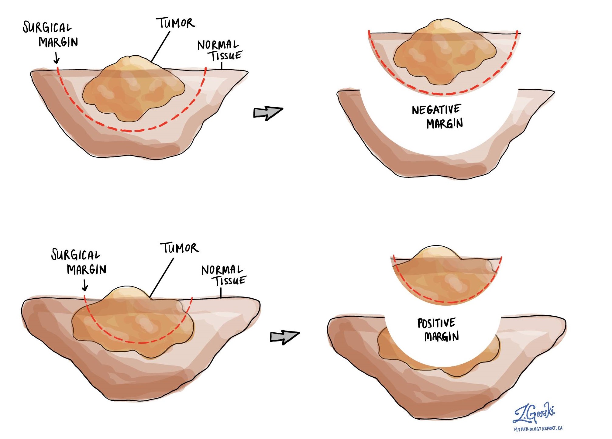
A margin is any tissue that has to be cut by a surgeon so that a tumour can be removed from your body. The appendix is connected to the large bowel, and generally has two areas that must be cut to remove it from the body. The end that attaches to the large bowel and is in direct communication with it is the proximal margin. The appendix has an area of fat that contains blood vessels and occasionally lymph nodes. This area must be cut to free the appendix and it is called the mesoappendix margin.
A negative margin means that no tumour cells or mucin were seen at the cut edge of the tissue. A positive margin means that your pathologist saw tumour cells or mucin at the cut edge of the tissue. A positive margin increases the risk that the tumour will regrow in the same location after surgery.

What does lymphovascular invasion mean and why is it important?
Lymphovascular invasion means that tumour cells were seen inside of a blood vessel or lymphatic vessel. Blood vessels are long thin tubes that carry blood around the body. Lymphatic vessels are similar to small blood vessels except that they carry a fluid called lymph instead of blood. Lymphovascular invasion is important because tumour cells can use blood vessels or lymphatic vessels to spread to other parts of the body such as lymph nodes or the lungs. However, it is extremely uncommon for tumour cells from a LAMN to show lymphovascular invasion.

Were lymph nodes examined and did any contain tumour cells?
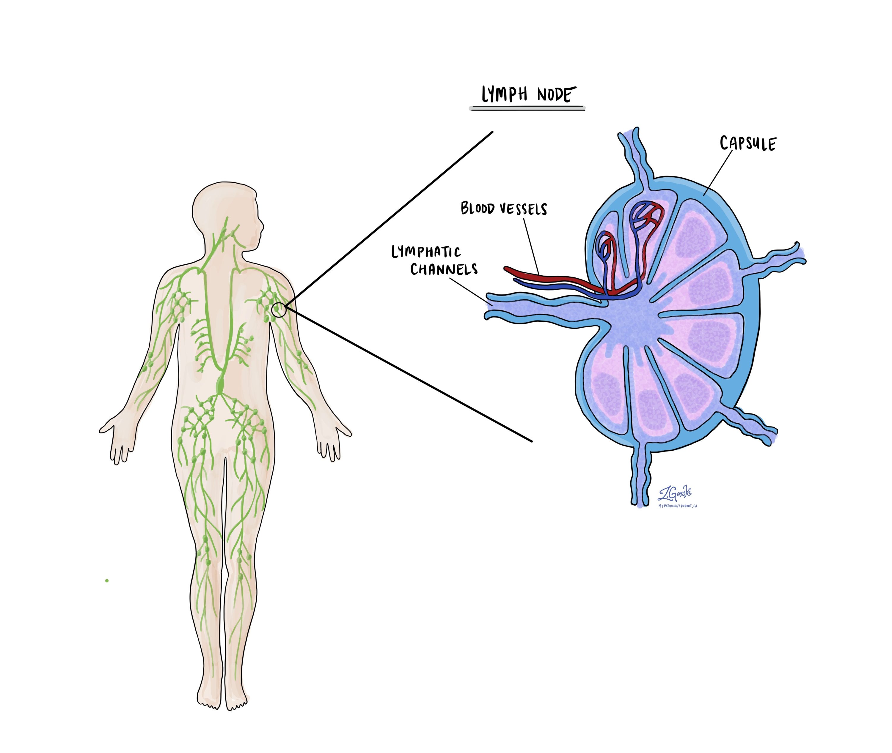
Lymph nodes are small immune organs located throughout the body. Tumour cells can spread from the tumour to a lymph node through lymphatic channels located in and around the tumour. The movement of tumour cells from the tumour to a lymph node is called a metastasis.
Your pathologist will carefully examine any lymph nodes removed with the tumour for tumour cells. Lymph nodes that contain tumour cells are often called positive while those that do not have any tumour cells are called negative. Most reports include the total number of lymph nodes examined and the number, if any, that have tumour cells. Lymph node metastases are important because they increase the risk that tumour cells will spread to other parts of the body. The examination of lymph nodes is also used to determine the pathologic nodal stage (pN). However, tumour cells from a LAMN rarely spread to lymph nodes.

What information is used to determine the pathologic stage for low grade appendiceal mucinous neoplasm?
The pathologic stage for LAMN is based on the TNM staging system, an internationally recognized system originally created by the American Joint Committee on Cancer. This system uses information about the primary tumour (T), lymph nodes (N), and distant metastatic disease (M) to determine the complete pathologic stage (pTNM). Your pathologist will examine the tissue submitted and give each part a number. In general, a higher number means a more advanced disease and a worse prognosis.
Tumour stage (pT)
LAMN is different from other tumours in the gastrointestinal tract because it does not have a T1 or T2 stage. The tumour stage (pT) for LAMN includes Tis, T3, T4a, and T4b.
- Tis (LAMN) – Tumour cells or the mucin they produce were only seen touching or invading the muscularis propria.
- T3 – Tumour cells or the mucin they produce were seen extending through the muscularis propria into the fat below the serosa (subserosa or mesoappendix).
- T4 – Tumour cells or the mucin they produce have broken through the serosa and are in the abdominal cavity. This category is further divided into T4a and T4b. In T4a, the tumour cells have broken through the serosa but are not seen on nearby organs. in T4b tumour cells are found on nearby organs.
Nodal stage (pN)
LAMN is given a nodal stage (pN) between 0 and 2. This is based on the number of lymph nodes that contain tumour cells. If there are no lymph nodes in the surgical specimen the nodal stage cannot be determined and it is listed as pNX. Lymph node involvement is extremely rare in low grade appendiceal mucinous neoplasms.
Metastasis stage (pM)
If tumour cells from a LAMN have spread throughout the abdominal space or into other organs away from the appendix it is considered to be metastatic and is given a metastatic stage of M1. This stage is then further divided into stages M1a, M1b, and M1c.
- M1a – This stage is given when there is mucin without tumour cells (acellular mucin) found in the abdominal cavity.
- M1b – This stage is given when there are tumour cells found in the abdominal cavity or on nearby organs.
- M1c – This stage is given if tumour cells are found outside the abdominal cavity.
The metastatic stage can only be determined if other organs, tissues, or mucin that was within the abdominal cavity are submitted to your pathologist for examination.


