

Parakeratosis is a word pathologists use to describe a change in the surface layer of squamous epithelium, which is the tissue that lines many parts of the body including the skin, mouth, throat, esophagus, cervix, and anal canal. In healthy tissue, the squamous cells at the surface mature and lose their nuclei (the part of the cell that holds genetic material) as they form a protective outer layer of keratin.
In parakeratosis, these surface cells retain their nuclei, which is not normally seen in mature keratin. This change usually means that the tissue is responding to irritation, inflammation, or injury. Parakeratosis is often seen in areas where the tissue is healing, regenerating, or under stress.
What causes parakeratosis?
Parakeratosis can be caused by a variety of conditions, most of which are non-cancerous.
It commonly occurs in tissues that are:
-
Inflamed or irritated, such as in chronic dermatitis or esophagitis.
-
Infected, including in fungal or viral infections.
-
Exposed to repeated trauma, such as friction, pressure, or chemical irritation.
-
Affected by underlying skin disorders, such as psoriasis or lichen planus.
-
Undergoing abnormal cell growth, such as dysplasia or cancer in rare cases.
Whether parakeratosis is a harmless change or something more concerning depends on the context, including other features seen in the tissue and the clinical history.
Is parakeratosis serious?
Parakeratosis on its own is not usually serious and is often a benign, reversible change. In most cases, it reflects normal tissue repair or a response to irritation.
However, when parakeratosis is seen alongside other abnormal findings—such as dysplasia (precancerous changes) or abnormal cell growth—it may be part of a more significant condition. Your doctor will interpret the finding of parakeratosis in combination with other parts of your pathology report and any relevant clinical information.
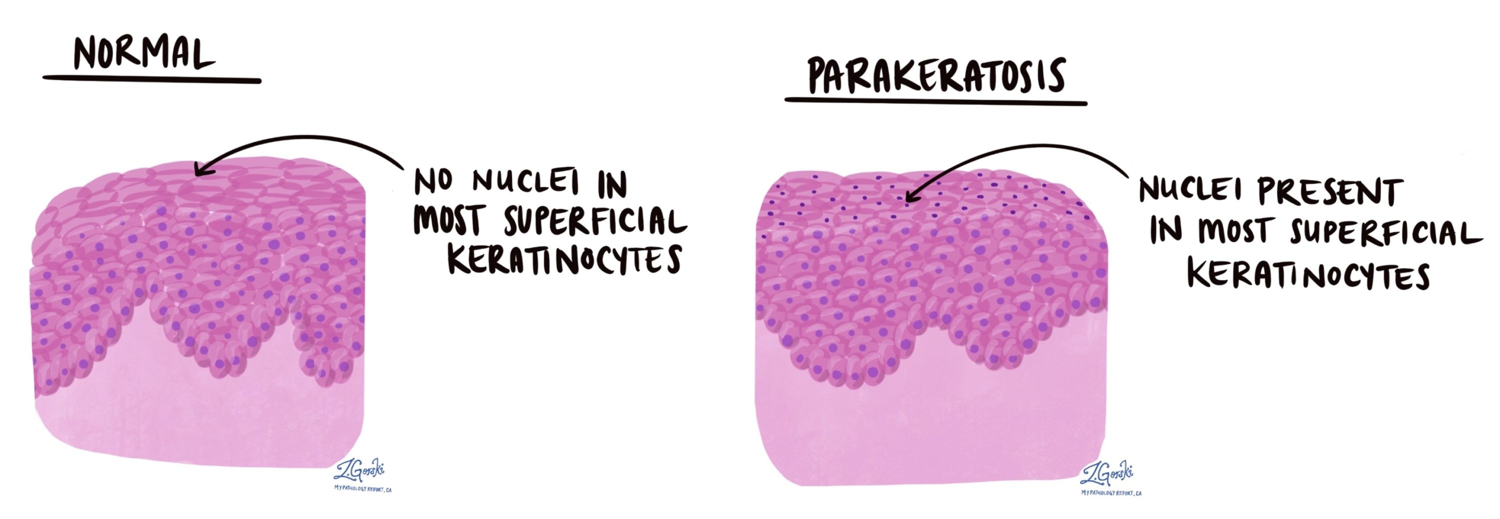
What does parakeratosis look like under the microscope?
Under the microscope, parakeratosis is seen as a thickened layer of keratin on the surface of squamous epithelium, with visible nuclei still present in the surface cells. This distinguishes it from orthokeratosis, where the surface cells have no nuclei, which is considered normal in many parts of the body.
Parakeratosis may be seen along with other surface changes such as:
-
Hyperkeratosis – a thicker-than-normal keratin layer,
-
Acanthosis – thickening of the entire squamous epithelium,
-
Inflammation – presence of immune cells in or under the epithelium,
Where in the body is parakeratosis commonly seen?
Parakeratosis can be seen in tissue from many different parts of the body lined by squamous epithelium, including:
-
Skin – commonly in conditions like psoriasis.
-
Esophagus – in reflux or irritation.
-
Cervix – in areas undergoing repair or with dysplasia.
-
Mouth and tongue – in response to friction or smoking.
-
Anal canal – especially in chronic irritation or HPV-related changes.
Questions to ask your doctor
-
What caused the parakeratosis in my case?
-
Are there any signs of dysplasia or cancer in my pathology report?
-
Do I need any follow-up or treatment?
-
Is this change likely to go away on its own?