

Acantholysis is a term pathologists use to describe the loss of normal connections between skin cells. In healthy skin, the cells of the outer layer — the epidermis — are held tightly together by specialized anchor structures called desmosomes, which act like tiny molecular rivets between cells. When desmosomes are damaged or destroyed, cells lose their grip on one another and begin to separate. This separation is called acantholysis. Depending on the cause and location, it can lead to visible blisters, erosions, or raw areas on the skin or the lining of the mouth and other mucous membranes.
What causes acantholysis?
Acantholysis is a finding, not a diagnosis in itself — it is a change that can be triggered by several different underlying conditions:
- Autoimmune blistering diseases — the most important cause to identify. In conditions such as pemphigus, the immune system produces antibodies that attack desmosomes, causing widespread cell separation and serious blistering. Identifying acantholysis in this context is critical because pemphigus requires specific immunosuppressive treatment.
- Viral infections — certain viruses, including herpes simplex virus and varicella (chickenpox), can damage the structures holding skin cells together, producing localized acantholysis and blister formation.
- Genetic disorders — rare inherited conditions such as Darier disease and Hailey-Hailey disease affect the proteins that regulate desmosome function, making the skin prone to repeated episodes of acantholysis.
- Cancer — acantholysis can be a feature of a specific variant of skin cancer called acantholytic squamous cell carcinoma, in which the cancer cells lose their normal attachments to each other. Recognising this pattern helps the pathologist correctly classify the tumour.
- Other inflammatory skin conditions — some reactive or inflammatory skin diseases can produce focal (limited) areas of acantholysis as part of a broader pattern of tissue change.
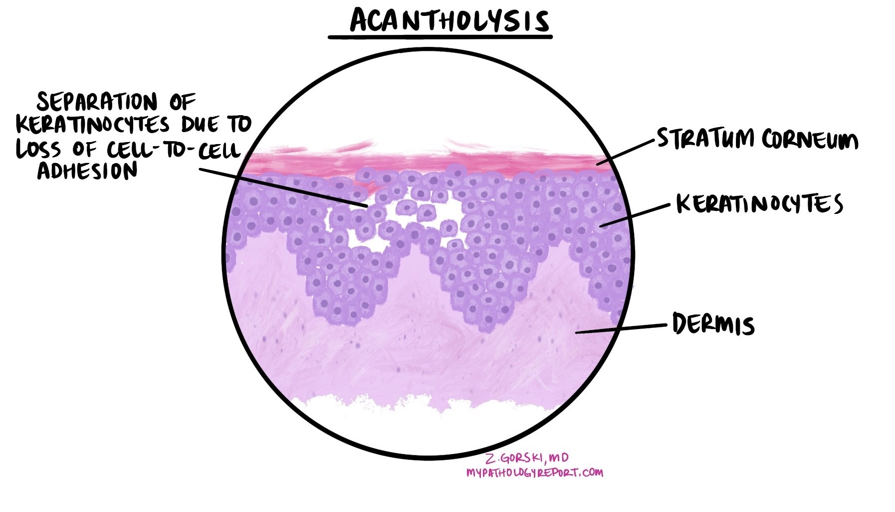
What does acantholysis look like under the microscope?
When a pathologist examines a skin biopsy showing acantholysis, the keratinocytes — the main cells of the epidermis — appear rounded and separated from each other, sometimes floating freely in spaces filled with fluid. This creates a characteristic “tombstone” appearance along the base of the epidermis in some types of pemphigus. The level within the skin layers at which the separation occurs is an important diagnostic clue: separation high in the epidermis suggests one type of pemphigus, while deeper separation points to another. The pathologist will note the location of the acantholysis and any other features — such as the presence of inflammatory cells, viral changes, or abnormal cancer cells — to help identify the underlying cause.
What does finding acantholysis in my report mean?
If your pathology report mentions acantholysis, it means the pathologist has identified an abnormal pattern of cell separation in the skin or mucous membrane sample. This finding is an important clue that helps narrow down the diagnosis, but the underlying cause still needs to be confirmed — usually with additional testing.
For suspected autoimmune blistering disease such as pemphigus, a test called direct immunofluorescence is typically performed on a separate skin sample. This test looks for antibodies deposited in the skin and can confirm the diagnosis and identify the specific type of blistering disease. Your dermatologist will guide you through any further steps needed.
Questions to ask your doctor
- What is the most likely cause of the acantholysis found in my biopsy?
- Will I need further tests — such as immunofluorescence — to confirm the diagnosis?
- What treatment is recommended based on this finding?
Related articles on MyPathologyReport.com
We are proud to partner with:
![]()