

Atrophic describes tissues or organs that have become smaller or thinner due to decreased cell size or number. This change is benign (non-cancerous) and reflects how the tissue appears under a microscope. It is not a specific medical diagnosis but is commonly used by pathologists to describe tissue changes.
What causes tissue to become atrophic?
Tissues can become atrophic for several reasons:
- Decreased blood supply: Organs require sufficient blood flow to stay healthy. When blood flow is reduced for a long time, tissues become atrophic. For example, tissues surrounding a large tumor may become atrophic due to pressure limiting blood supply.
- Disuse (lack of use): Tissues or muscles that aren’t regularly used can become atrophic. Muscle shrinkage after prolonged inactivity or injury is a typical example.
- Decreased hormone stimulation: Certain organs depend on hormones to maintain size and function. With age, hormone levels decrease, causing organs such as the breast and uterus lining (endometrium) to become atrophic.
- Chronic inflammation: Long-term inflammation can damage tissues, leading to atrophic changes. A typical example is atrophic gastritis, where persistent inflammation of the stomach lining causes the tissue to thin.
Where is atrophic tissue commonly found?
Atrophic changes can occur anywhere in the body but are most commonly seen in:
- Muscles after periods of inactivity.
- Breast tissue and uterus lining after menopause.
- Brain tissue in older adults.
- Tissues are affected by poor blood flow.
- The stomach, particularly in conditions like atrophic gastritis.
What are the symptoms of atrophic tissue?
Symptoms depend on the location:
- Muscle atrophy can cause weakness or visible shrinking.
- Breast or uterine tissue typically shows no symptoms but may lead to physical changes.
- Brain atrophy can cause memory loss or difficulty thinking.
- Atrophic gastritis can lead to digestive discomfort or nutrient deficiencies.
Many times, especially in internal organs, atrophic changes cause no symptoms and may only be discovered during medical exams.
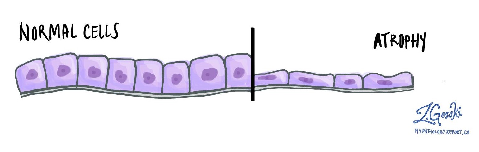
What does atrophic tissue look like under the microscope?
Under the microscope, atrophic tissue appears thinner than normal, with smaller cells and larger spaces between them. These microscopic changes indicate the tissue has adapted to decreased use, limited blood flow, reduced hormone stimulation, or ongoing inflammation.
We are proud to partner with:
![]()