

Atypical mitosis is an abnormal cell division. Cells normally copy themselves through a tightly controlled process called mitosis, which evenly divides the cell’s genetic material (DNA) so that each new cell receives a complete and matching set. In an atypical mitosis, this process goes wrong and the DNA is not shared evenly. This is the same finding that pathologists also call an atypical mitotic figure, the term for a dividing cell with this abnormal appearance under the microscope. This article explains what atypical mitosis means and why it may appear in your pathology report.
What does atypical mitosis look like under the microscope?
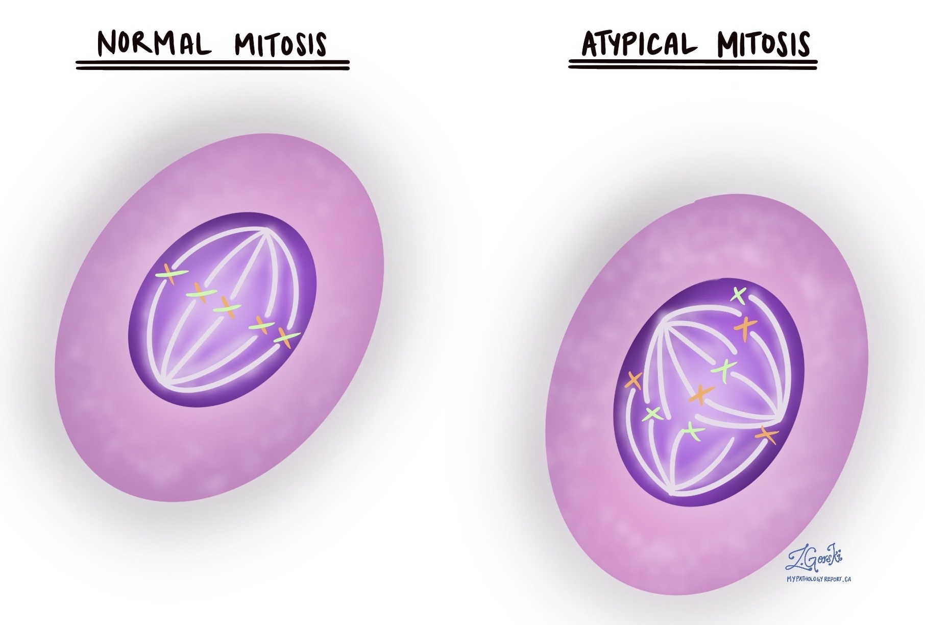
In a normal cell division, the chromosomes (the structures that carry DNA) gather in the middle of the cell and are evenly pulled toward opposite ends, producing two new cells with identical sets of DNA. An atypical mitosis looks different because the orderly steps have broken down. The chromosomes may be pulled toward three or more directions at once (described as tripolar or multipolar), arranged in a ring, or left scattered and disorganized. Because the division is uneven, the new cells end up with too much, too little, or jumbled genetic material.
Why is atypical mitosis important?
Atypical mitosis is important because it indicates that cells are dividing in an uncontrolled, disorderly way, a feature of cancer. Uneven DNA sharing produces cells with an abnormal amount of genetic material, and this kind of genetic instability is one of the ways cancers develop and change over time. Because atypical mitoses are uncommon in normal tissue, finding them is a helpful clue that supports recognition and diagnosis of cancer. They also contribute to a tumor’s grade, a measure of how abnormal the cells look and how quickly they appear to be growing.
Does atypical mitosis always mean cancer?
Not by itself. Atypical mitoses point strongly toward cancer, but they are not enough on their own to make the diagnosis. Abnormal divisions can occasionally appear in tissue damaged by radiation therapy, chemotherapy, or certain infections, and rarely in some noncancerous conditions. They are also most often seen in cancers such as breast, lung, and colon cancer and melanoma, and are usually absent from healthy tissue. For these reasons, a pathologist always weighs this finding alongside the other features of the tissue and sometimes additional tests before reaching a final diagnosis.
What happens after atypical mitosis is found?
When atypical mitoses are identified, the pathologist examines them alongside the other microscopic features of the tissue and may order additional tests, such as immunohistochemistry (IHC), to determine the type and origin of the abnormal cells. Immunohistochemistry uses markers that highlight specific proteins to help confirm what kind of cells are present. Together, these findings contribute to the final diagnosis and, when cancer is confirmed, to the tumor’s grade. The diagnosis, grade, type, and stage are what the care team uses to plan next steps and discuss treatment options, rather than the finding of atypical mitosis alone.
Questions to ask your doctor
- What does the finding of atypical mitosis mean in my case?
- Was atypical mitosis part of how my diagnosis was made?
- Did this finding affect the grade of my tumor?
- Were any additional tests, such as immunohistochemistry, performed on my sample?
- Could this finding be related to previous radiation or chemotherapy rather than cancer?
- What do the diagnosis and grade tell us about how this condition is likely to behave?
- What are the next steps based on my overall pathology results?
We are proud to partner with:
![]()