

Cautery artifact is a term pathologists use to describe changes in the appearance of cells and tissue caused by heat during a surgical procedure. These changes are not related to disease — they are a side effect of a common surgical tool called electrocautery, which uses electrical heat to cut tissue and seal bleeding blood vessels. Electrocautery is used routinely during biopsies and surgeries, and the heat it generates can alter the appearance of the tissue at and near the edges of the removed specimen. When a pathologist notes cautery artifact in your report, it is an observation about tissue handling — not a new finding related to your diagnosis.
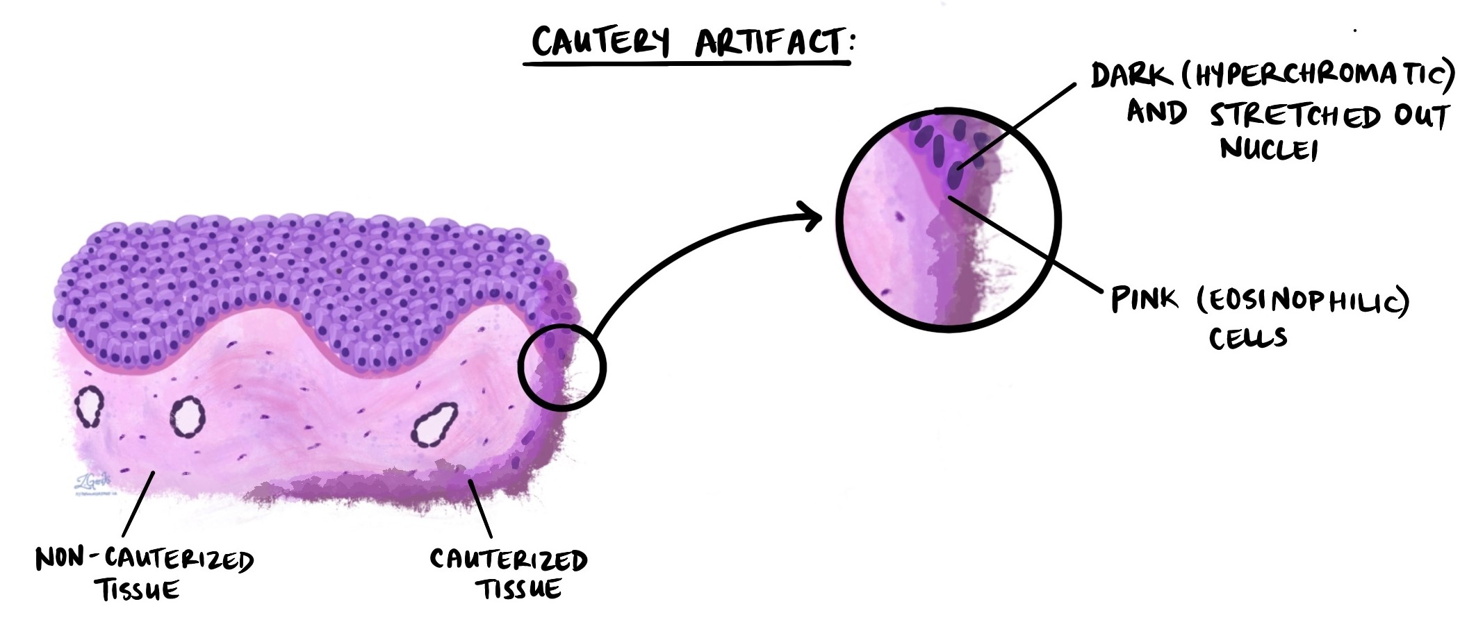
What does cautery artifact look like under the microscope?
When tissue affected by cautery is examined under the microscope, the heat-damaged cells show characteristic changes that distinguish them from disease-related abnormalities:
- Dense, pink cytoplasm — the body of the cell (cytoplasm) becomes compacted and stains a brighter, deeper pink than normal, reflecting protein denaturation from the heat.
- Elongated or irregular nuclei — the nuclei — the part of each cell that contains DNA — may appear stretched, smeared, darkened, or irregular in shape. These changes can superficially resemble the abnormal nuclei seen in cancer or precancerous conditions.
- Loss of cellular detail — cells may appear shrunken, blurred, or fragmented, making it difficult to assess their true nature.
Because these heat-related changes can mimic disease, experienced pathologists are trained to recognize cautery artifact and distinguish it from genuine abnormality. The artifact is typically most pronounced at the edges of the specimen and becomes less prominent in areas farther from the cut margin.
Why does cautery artifact matter in a pathology report?
In most cases, cautery artifact is a minor finding that does not affect the overall diagnosis — there is usually enough unaffected tissue in the sample for the pathologist to make a clear assessment. However, when the artifact involves a critical area of the specimen, it can pose challenges:
- Diagnosis — if the most suspicious or representative area of the tissue is heavily affected by cautery, the pathologist may not be able to confirm whether the cells are normal, precancerous (dysplasia), or malignant.
- Surgical margins — one of the most important questions a pathologist answers is whether a tumor was completely removed. The surgical margin is the edge of the removed tissue, and the pathologist examines it closely to confirm no cancer cells remain at the cut edge. When cautery artifact obscures the margin, it may be impossible to determine with confidence whether the margin is clear or involved.
When cautery artifact affects a critical area, the pathologist will note in the report that evaluation was limited and may recommend clinical correlation or additional testing.
What does cautery artifact mean for my diagnosis and treatment?
In most cases, finding cautery artifact in your report does not change your diagnosis or treatment. It is a routine observation that reflects how the tissue was removed, not something that indicates a problem with your care. However, if the artifact affects the surgical margins or the area being evaluated for cancer, your doctor may recommend further steps — such as a repeat biopsy, re-excision, or additional imaging — to ensure a complete and accurate assessment.
If you are uncertain whether cautery artifact has affected the interpretation of your results, it is entirely appropriate to ask your doctor or pathologist for clarification.
Questions to ask your doctor
- Did cautery artifact affect any critical areas of my sample — such as the surgical margins or a suspicious lesion?
- Is the pathologist confident in the diagnosis despite the artifact, or is further testing needed?
- Do I need a repeat biopsy or re-excision based on this finding?
Related articles on MyPathologyReport.com
We are proud to partner with:
![]()