

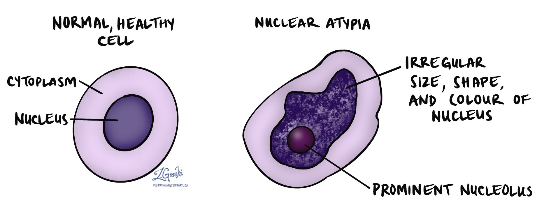
In a pathology report, the term nuclear atypia is used to describe cells that have abnormal-looking nuclei when examined under a microscope. The nucleus is the part of the cell that contains its genetic material (DNA) and controls how the cell functions. Pathologists use the word “atypia” to mean that something looks unusual or different from normal.
A nucleus may be described as atypical if it is:
-
Larger than normal.
-
Darker than normal (this is called hyperchromatic).
-
Irregular in shape.
-
Has clumped or coarse chromatin (the DNA material inside the nucleus appears unevenly distributed).
-
Contains prominent nucleoli, which are round structures within the nucleus that can become large or more visible in abnormal cells.
Nuclear atypia is a descriptive term, not a diagnosis on its own. It simply means the pathologist noticed that the cells are not completely normal in appearance.
What causes nuclear atypia?
Nuclear atypia can result from changes inside the cell or from external factors that affect the tissue. Some causes include:
-
Genetic mutations – Abnormal changes in the DNA can cause cells to grow and divide in an uncontrolled way. This can lead to pre-cancerous changes or cancer over time.
-
Inflammation – Cells in inflamed tissue may appear atypical due to stress or injury.
-
Viral infections – Certain viruses (like HPV or EBV) can cause cells to look abnormal.
-
Radiation exposure – Previous radiation therapy or accidental exposure can damage cells and cause nuclear changes.
-
Medications or treatments – Some drugs, especially chemotherapy, can cause nuclear atypia in treated tissues.
-
Tissue damage – Atypia may be seen in tissues that have been injured due to trauma, lack of blood flow, or healing.
Does nuclear atypia mean cancer?
No. Nuclear atypia does not mean cancer. While it is true that many cancers show nuclear atypia, not all cells with atypical nuclei are cancerous.
-
In benign (non-cancerous) conditions, nuclear atypia may be mild and related to inflammation, infection, or healing.
-
In precancerous conditions, nuclear atypia may be more noticeable and may indicate a higher risk of cancer developing over time.
-
In cancer, nuclear atypia is often more severe, with very large, dark, and irregular nuclei, and high rates of cell division.
Because nuclear atypia can occur in both non-cancerous and cancerous situations, the context is very important. Pathologists consider the pattern, severity, and other features of the tissue when deciding what the nuclear atypia means.
How do pathologists describe nuclear atypia?
Pathologists often use additional words to describe the degree of atypia:
-
Mild atypia – Small changes in the size or shape of the nucleus; may be seen in reactive or inflamed tissue.
-
Moderate atypia – More noticeable changes that may raise concern, especially if found in high numbers.
-
Severe atypia – Marked nuclear changes that may suggest a precancerous or cancerous condition.
These descriptions help guide further testing, follow-up, or treatment.
What happens if nuclear atypia is found?
If nuclear atypia is seen in a tissue sample, your doctor will consider:
-
The location of the abnormal cells
-
The underlying cause (such as inflammation, infection, or tumour)
-
Whether the atypia is mild or severe
-
Whether the atypical cells are limited to a small area or widespread
-
Whether other features of cancer are present (such as invasion into surrounding tissue)
Depending on these factors, your doctor may recommend:
-
No further treatment, if the atypia is mild and related to a benign condition.
-
Repeat testing or closer monitoring over time.
-
Removal of the tissue if the atypia is suspicious or occurs in a high-risk area.
-
Further testing such as immunohistochemistry or molecular analysis if cancer is suspected.
Questions to ask your doctor
-
What does the nuclear atypia mean in my pathology report?
-
Is the atypia related to inflammation, infection, or something else?
-
Is this a sign of a precancerous or cancerous change?
-
Do I need further tests or follow-up?
-
What are the next steps in my care?
We are proud to partner with:
![]()