

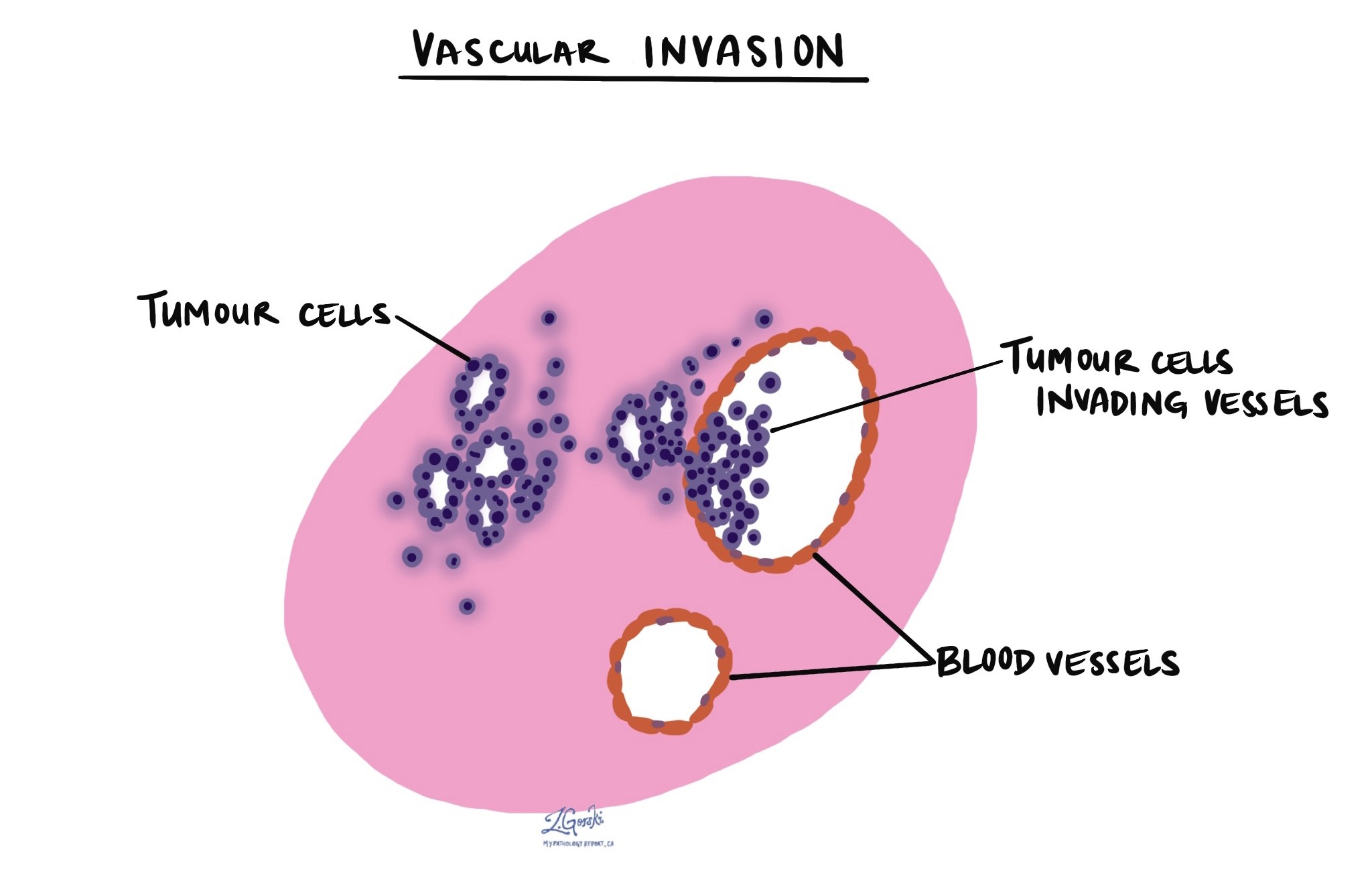
Vascular invasion means that cancer cells have grown into a blood vessel. Pathologists also call it angioinvasion. Blood vessels carry blood through the body, so cancer cells that enter them can travel to other organs. A pathologist looks for vascular invasion when examining a tumor under the microscope.
Vascular invasion is a finding, not a diagnosis. It describes what the pathologist saw in the tissue. This article explains what vascular invasion means, how pathologists identify it, what the wording on your report means, and why the finding matters.
What does vascular invasion mean?
A growing tumor can invade the surrounding tissue. Vascular invasion happens when tumor cells push into the wall of a blood vessel or enter the space inside it. Cancer cells most often invade veins. Veins have thinner walls than arteries, so cells enter them more easily.
Once cancer cells are inside a vein, the blood can carry them away from the tumor. This is how some cancers metastasize, or spread to distant organs. Blood from the bowel flows to the liver first, so bowel cancers often spread there. Blood from most other organs flows to the lungs first.
How is vascular invasion different from lymphovascular invasion?
Pathologists use several related terms for cancer cells inside vessels. Reports use them in different ways:
- Vascular invasion. Tumor cells are inside a blood vessel. This term usually refers to veins. Some reports call it venous invasion or angioinvasion.
- Lymphatic invasion. Tumor cells are inside a lymphatic vessel. These thin channels carry fluid to the lymph nodes. See lymphatic invasion.
- Lymphovascular invasion. This broader term covers both types of vessels. Reports often use it when the pathologist cannot tell a small blood vessel from a lymphatic vessel. See lymphovascular invasion.
Some reports separate small-vessel invasion from large-vessel invasion. Small vessels are thin-walled channels that can be lymphatics or tiny blood vessels. Large vessels are veins with a muscular wall. Your report may list each one separately.
How is vascular invasion identified?
The pathologist identifies vascular invasion by examining thin slices of the tumor and surrounding tissue under the microscope. The pathologist looks for a cluster of tumor cells within a space lined by the wall of a vessel. Veins have a muscular wall, and some contain red blood cells.
The finding can be hard to confirm. Tissue can shrink during processing, leaving a gap around a group of tumor cells. This gap, called retraction, can look like a vessel. To avoid a mistake, the pathologist may use extra tests:
- Immunohistochemistry. immunohistochemistry uses antibodies to mark the cells that line vessels. CD31, CD34, and ERG mark blood vessels. D2-40 marks lymphatic vessels.
- Special stains. An elastic special stain outlines the elastic layer in a vein wall. This helps the pathologist confirm that a space is a vein.
What might my report say about vascular invasion?
Reports state the result in a few standard ways. The wording below is common, but each laboratory phrases it a little differently:
- Present, identified, or positive. The pathologist saw tumor cells inside a blood vessel. This is the finding described in this article.
- Not identified, absent, or negative. The pathologist did not see tumor cells in a blood vessel in the tissue examined. The pathologist checks only the slides prepared from the tumor, so this result lowers the concern but cannot rule out invasion.
- Indeterminate or suspicious. Something looked like vascular invasion, but the pathologist could not be sure. Retraction around tumor cells is a common cause.
- Extramural or intramural. In bowel cancers, the report may specify which vein was invaded. Extramural means outside the muscle wall of the bowel. Intramural means within the wall.
Does vascular invasion mean the cancer has spread?
Vascular invasion does not mean the cancer has spread. It means the cancer has a route to spread, and the chance of spread is higher. Many people with vascular invasion never develop spread, and some people without it do. Imaging tests and other results show whether cancer has reached other organs.
Why is vascular invasion important?
Doctors consider vascular invasion an adverse feature in most cancers. The finding matters in four ways:
- Risk of spread. Tumor cells in a blood vessel can reach distant organs. This raises the chance that the cancer will come back or spread.
- Prognosis. In many cancers, vascular invasion is linked to a worse prognosis. In colorectal cancer, invasion of veins outside the bowel wall predicts outcome independently of the stage.
- Staging. In some cancers, vascular invasion changes the stage. The AJCC 8th edition staging system for hepatocellular carcinoma uses vascular invasion. In a single tumor larger than 2 cm, it moves the tumor stage from T1b to T2. The TNM system also lets pathologists record venous invasion as a separate category.
- Diagnosis. In a few tumors, the number of invaded vessels helps classify the tumor. This is true for follicular thyroid carcinoma, where extensive vascular invasion carries a higher risk than limited invasion.
How does vascular invasion affect treatment?
Vascular invasion does not decide treatment on its own. The care team weighs it with the tumor type, grade, stage, and other findings in your report. In some cancers, vascular invasion leads the team to consider additional treatment after surgery or closer follow-up. Your doctor can explain how the finding applies to your situation.
Questions to ask your doctor
- Did the pathologist find vascular invasion in my tumor?
- Does my report separate blood vessel invasion from lymphatic invasion?
- Was the invaded vessel a small vessel or a large vein?
- If the result is indeterminate, what made it uncertain?
- Were special stains or immunohistochemistry used to confirm the finding?
- Does vascular invasion change my cancer stage?
- Does this finding affect my treatment options or follow-up?
- What tests will show whether the cancer has spread?
Related articles on MyPathologyReport.com
We are proud to partner with:


🔍 Search MyPathologyReport
Type what you see on your report — for example a diagnosis or test name