

Vasculitis means inflammation of blood vessel walls. It can affect arteries, veins, and capillaries. The inflammation can weaken, narrow, or block a vessel. Vasculitis is not one disease. It is a group of conditions that share this pattern of damage.
On a pathology report, vasculitis describes what the pathologist saw under the microscope. Your care team combines it with your symptoms and test results to name the specific type. This article explains what vasculitis is, how doctors classify it, how pathologists identify it, and what the wording on your report means.
What is vasculitis?
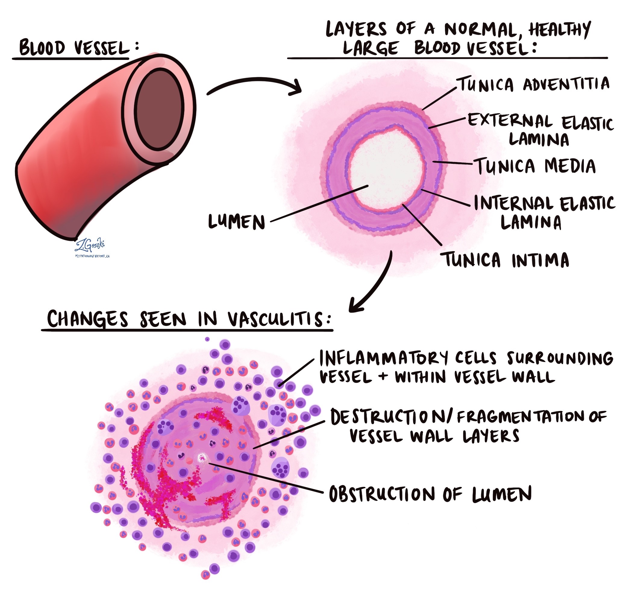
Vasculitis is inflammation in the wall of a blood vessel. Blood vessels carry blood to and from every organ. The heart and blood vessels article describes them in more detail. In vasculitis, inflammatory cells move into the vessel wall. The wall swells and thickens, and the cells can damage it.
A damaged vessel can narrow or close. The tissue it feeds then receives less blood. In severe cases, the vessel can bleed, form a clot, or rupture. The severity ranges from mild to life-threatening. It depends on which vessels and which organs are involved.
What are the symptoms of vasculitis?
Vasculitis symptoms vary widely because the disease can affect many organs. Common symptoms include fever, tiredness, muscle and joint pain, and weight loss. Skin involvement causes rashes or ulcers. Nerve involvement causes numbness or tingling. Lung involvement causes breathing difficulty or chest pain. Kidney involvement causes kidney problems. Vasculitis in the head can cause headache, confusion, or vision problems.
What causes vasculitis?
The cause of vasculitis is often unknown. In many cases, the immune system mistakenly attacks the vessel wall. Doctors call this an autoimmune process. Some forms of vasculitis have a known trigger or link:
- Autoimmune diseases. Vasculitis can occur in people with lupus, rheumatoid arthritis, or sarcoidosis.
- Infections. Doctors link hepatitis B, hepatitis C, and syphilis to some forms.
- Medicines. Some medicines can trigger vasculitis. Your doctor decides whether to change a medicine.
- Cancer. Vasculitis is rarely linked to cancer.
How do doctors classify vasculitis?
Doctors classify vasculitis mainly by the size of the vessels involved. The 2012 revised International Chapel Hill Consensus Conference nomenclature is the most widely used system. It sorts primary vasculitis into these groups:
- Large vessel vasculitis. This group affects the largest arteries. It includes Takayasu arteritis and giant cell arteritis.
- Medium vessel vasculitis. This group includes polyarteritis nodosa and Kawasaki disease.
- Small vessel vasculitis. This group has two parts. ANCA-associated vasculitis includes microscopic polyangiitis, granulomatosis with polyangiitis (formerly Wegener granulomatosis), and eosinophilic granulomatosis with polyangiitis (formerly Churg-Strauss syndrome). Immune complex vasculitis includes anti-glomerular basement membrane disease, cryoglobulinemic vasculitis, IgA vasculitis (formerly Henoch-Schönlein purpura), and hypocomplementemic urticarial vasculitis.
- Variable vessel vasculitis. This group can affect vessels of any size. It includes Behçet disease and Cogan syndrome.
- Single-organ vasculitis. This group affects one organ only. Examples include cutaneous leukocytoclastic angiitis, cutaneous arteritis, primary central nervous system vasculitis, and isolated aortitis.
The system also lists two secondary groups. One group occurs with another disease, such as lupus or rheumatoid arthritis. The other group has a probable cause, such as hepatitis C infection, a medicine, or cancer.
What does vasculitis look like under the microscope?
Under the microscope, vasculitis shows inflammatory cells in or around the vessel wall. The cell type and pattern of damage help the pathologist narrow the diagnosis. Three patterns are common:
- Neutrophil-rich pattern. Neutrophils fill and surround the vessel wall. Some of them break down and leave fragments called nuclear dust. Red blood cells may leak into the tissue. Doctors call this pattern leukocytoclastic vasculitis. It often affects small blood vessels in the skin. A higher number of eosinophils may point to a medicine as the cause.
- Granulomatous pattern. In giant cell arteritis, the inflammation forms granulomas that often contain giant cells. It spreads through all layers of the vessel wall and often damages the inner elastic layer. Some small-vessel vasculitides also cause necrotizing granulomatous inflammation.
- Eosinophil-rich pattern. Eosinophilic granulomatosis with polyangiitis shows eosinophil-rich inflammation. It also shows granulomas and often affects the airways.
The pathologist may also see fibrinoid necrosis. This is dead tissue in the vessel wall, mixed with a protein called fibrin. It is not always visible. Findings also change with biopsy timing, so that an early lesion may look different from an older one.
How is vasculitis diagnosed?
Doctors diagnose vasculitis by combining several types of information. They review your symptoms and perform an exam. They order blood tests that measure inflammation and look for autoantibodies. They often order imaging. Doctors often take a biopsy of an affected organ, such as the skin, a temporal artery, or the kidney.
The laboratory may add special tests to the biopsy:
- Direct immunofluorescence. Direct immunofluorescence uses fluorescent antibodies to find immune proteins in the vessel wall. In skin, it can show fibrinogen, C3, IgG, and IgM. IgA suggests IgA vasculitis, but IgA also appears in other conditions. A biopsy of an affected skin area yields more.
- Kidney biopsy. Some vasculitides damage the kidney filters. The pathologist may find a pauci-immune pattern, which means few or no immune deposits. The pathologist may also find crescents, which are crescent-shaped collections of cells around the filter, and fibrinoid necrosis.
- ANCA blood tests. ANCA stands for antineutrophil cytoplasmic antibody. Two types, MPO and PR3, are common in ANCA-associated vasculitis. The ANCA type alone does not provide a specific diagnosis. About one in five people with this kidney pattern have no ANCA in their blood.
What might my report say about vasculitis?
Reports describe vasculitis in a few standard ways. Each laboratory phrases it a little differently:
- Vasculitis identified. The pathologist saw inflammation in a vessel wall. The report may describe the vessel size, such as small-vessel or medium-vessel.
- Leukocytoclastic vasculitis. The inflammation is rich in neutrophils and nuclear dust.
- Necrotizing vasculitis. The vessel wall contains areas of dead tissue.
- Granulomatous vasculitis or arteritis. The inflammation forms granulomas.
- No vasculitis identified. The pathologist did not see vasculitis in the sample. A biopsy removes only a small piece of tissue, so this result lowers concern but cannot rule out vasculitis.
- A comment about clinical correlation: the pathologist asks your doctor to compare the biopsy with your symptoms and test results.
Is vasculitis cancer?
Vasculitis is not cancer. It is inflammation of blood vessels, not abnormal cell growth. Cancer is a rare cause of vasculitis in some people, and your doctor can tell you whether that applies to you.
What happens after vasculitis is identified?
The next steps depend on the type of vasculitis and the organs involved. Your care team may include specialists, such as a rheumatologist. Doctors may order more blood tests, imaging, or another biopsy to see how widely the disease has spread. Your doctor can explain the treatment plan for your type of vasculitis.
Questions to ask your doctor
- What type of vasculitis does my biopsy show?
- Does the vasculitis involve small, medium, or large vessels?
- Does my report mention necrosis, granulomas, or eosinophils?
- Was immunofluorescence done, and what did it show?
- Which organs could vasculitis affect?
- Do I need blood tests such as ANCA?
- Could a medicine, an infection, or an autoimmune disease be the cause?
- Do I need another biopsy or imaging?
- Which specialist will manage my care?
Related articles on MyPathologyReport.com
We are proud to partner with:


🔍 Search MyPathologyReport
Type what you see on your report — for example a diagnosis or test name