

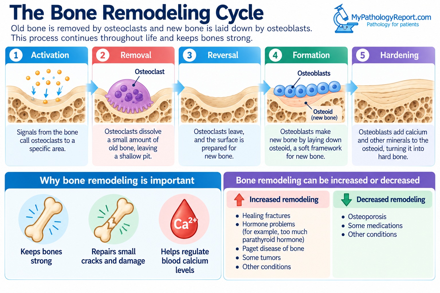
Bone remodeling is the process the body uses to renew its bones constantly. Old or damaged bone is removed by cells called osteoclasts, and new bone is laid down in its place by cells called osteoblasts. This process continues throughout life. It keeps bones strong, repairs small cracks, and helps regulate blood calcium levels.
Bone remodeling is a normal process, not a diagnosis. Pathology reports may mention it when a bone sample shows more or less remodeling than expected. This article explains how bone remodeling works, what it looks like under the microscope, and why it may be mentioned in your report.
How does bone remodeling work?
Bone remodeling happens in small areas spread throughout the skeleton. Each area goes through the same series of steps:
- Activation. Signals from the bone, often in response to wear and tear, call osteoclasts to a particular area.
- Removal. Osteoclasts dissolve a small amount of old bone, leaving a shallow pit.
- Reversal. Osteoclasts leave, and the surface is prepared for new bone.
- Formation. Osteoblasts fill the pit with osteoid, the soft material that forms the framework of new bone.
- Hardening. Osteoblasts add calcium and other minerals to the osteoid, turning it into hard bone.
Cells called osteocytes, which live inside the bone, sense stress and damage. They help decide where remodeling is needed.
What controls bone remodeling?
Several factors control the balance between bone removal and bone formation. These include parathyroid hormone, vitamin D, and blood calcium levels. Sex hormones such as estrogen and testosterone help protect bone, and physical activity encourages new bone to form. Some medications can also affect bone remodeling.
When removal and formation are balanced, bone stays strong. When removal outpaces formation, bone becomes thinner and weaker over time.
What does bone remodeling look like under the microscope?
Under the microscope, a pathologist can see signs of bone remodeling on the bone surface. Osteoclasts sit in small, scooped-out pits where they remove bone. Rows of osteoblasts line areas where new bone is being made, often on top of a pale layer of osteoid.
The pathologist can also see the type of bone. Newly made bone, called woven bone, has a disorganized structure. Mature bone, called lamellar bone, is arranged in neat parallel layers. Large amounts of woven bone indicate rapid bone formation.
Why is bone remodeling mentioned in a pathology report?
Changes in bone remodeling can indicate several conditions. Your report may describe remodeling as “increased,” “active,” or “reactive.” Common reasons include:
- Healing fractures. After a fracture, remodeling increases sharply as the body repairs the bone. Healing bone can look very active under the microscope.
- Hormone problems. Too much parathyroid hormone, often caused by a parathyroid adenoma, increases bone removal.
- Osteoporosis. In osteoporosis, bone is removed faster than it is replaced, leaving thin, weak bone.
- Paget disease of bone. In this condition, remodeling is extremely fast and disorganized, producing large but weak bones.
- Tumors. Many tumors in or near bone, including cancer that has spread to bone, cause the surrounding bone to remodel.
Several medicines used for bone conditions slow bone removal. Blood tests, such as parathyroid hormone and calcium tests, can help identify the cause of abnormal remodeling.
Questions to ask your doctor
- Why does my report mention bone remodeling?
- Is the remodeling in my sample increased or decreased?
- Does the remodeling suggest a healing fracture, a hormone problem, or another cause?
- Should my calcium, vitamin D, or parathyroid hormone levels be checked?
- Do I need a bone density test?
- What is the final diagnosis in my report?
- Do I need any further treatment or follow-up?
Related articles on MyPathologyReport.com
We are proud to partner with:


🔍 Search MyPathologyReport
Type what you see on your report — for example a diagnosis or test name