

Stroma is the supporting tissue that forms the framework of an organ. It holds the organ together and gives it strength and structure, while also carrying the blood vessels and other channels the organ needs. Pathologists contrast the stroma with the parenchyma, which is the working part of the organ that does its main job, such as the epithelial cells that line a gland. Every organ has stroma, although its appearance and thickness vary from one part of the body to another.
Stroma is a normal part of the body, and the word appears often on pathology reports because the pathologist examines both the working cells of a tissue and the stroma around them. This article explains what stroma is made of, how it changes in response to injury and cancer, and why it matters when preparing a report.
What is stroma made of?
Stroma is a type of connective tissue. It consists of a network of thin supporting fibers, the cells that produce them, and the blood and lymphatic vessels that run through it. Its main components are:
- Fibroblasts — Long, thin cells, often described as spindle-shaped, that produce the fibers giving the stroma its strength.
- Supporting fibers — A mesh of fibers, mainly a protein called collagen, that forms a stable framework for the organ.
- Blood and lymphatic vessels — Blood vessels deliver oxygen and nutrients, and lymphatic channels help remove excess fluid and waste.
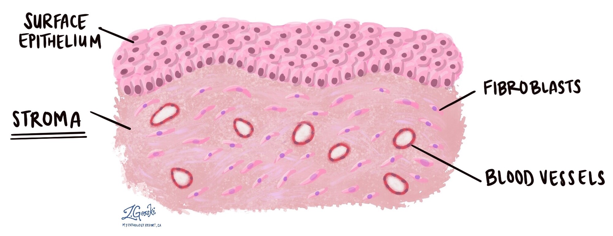
What does the stroma look like under the microscope?
Under the microscope, normal stroma appears as a network of thin fibers with scattered spindle-shaped fibroblasts, forming a stable background that supports the organ’s working cells. Blood vessels and lymphatic channels run through it. The stroma can change in response to injury, inflammation, or cancer, and pathologists pay close attention to these changes because they provide clues about what is happening in the surrounding tissue.
How does the stroma change in response to cancer?
When cancer cells invade a previously normal area, the stroma often reacts. A common reaction is called desmoplasia, in which the stroma becomes dense, fibrous, and scar-like. Under the microscope, it looks thickened and firm. Desmoplasia can help the pathologist recognize that a tumor is invasive, because noncancerous growths usually do not trigger this kind of stromal reaction. Desmoplasia is seen in many cancers, including those of the breast, colon, pancreas, and stomach.
What is reactive stroma?
Not all stromal changes are caused by cancer. Reactive stroma is the term pathologists use for noncancerous stromal changes that occur when tissue responds to injury or stress. It may appear in tissue surrounding a tumor, in areas healing after surgery or radiation therapy, and at sites of inflammation or trauma. Although reactive stroma can look different from normal stroma, it does not mean cancer is present. Instead, it reflects the body’s attempt to repair or respond to an abnormal condition. Telling reactive stromal changes apart from a true cancer-related reaction is one of the routine judgments a pathologist makes.
Why does the stroma matter when assessing a biopsy for cancer?
One of the most important questions in assessing a biopsy for cancer is whether a tumor is invasive. Invasion means that abnormal cells have moved beyond their normal location into the surrounding stroma. This distinction matters because an invasive cancer is managed differently from a noninvasive one, such as a carcinoma in situ, in which the abnormal cells have not yet crossed into the stroma. To determine whether invasion is present, the pathologist needs to see both the abnormal cells and the surrounding stroma.
What happens when a biopsy does not contain stroma?
Some biopsies sample only the surface of an abnormal area and miss the underlying stroma. In that case, the pathologist may not be able to determine whether the tumor is invasive. The report may then say something like “cannot exclude invasion” or “at least in situ,” meaning the sample does not allow full assessment of invasion. In this situation, the pathologist may recommend more tissue, such as a deeper biopsy or a larger removal, to complete the evaluation.
Missing stroma in a biopsy does not mean the tumor is more advanced or faster-growing. It means the sample is incomplete, and more tissue is needed to make a precise diagnosis.
Why is the stroma important in a pathology report?
Stroma changes help the pathologist understand how a tumor is behaving. The presence of desmoplasia may support a diagnosis of invasive cancer, while the absence of a stromal reaction may support a noninvasive or noncancerous condition. Reactive stromal changes can also help explain findings around a tumor or in tissue affected by injury or treatment. By examining the stroma alongside the tissue’s working cells, the pathologist gathers information that contributes to diagnosis, treatment decisions, and prognosis.
Questions to ask your doctor
- What changes, if any, were seen in the stroma of my tissue sample?
- Was there enough stroma present to assess whether the tumor is invasive?
- Do I need another biopsy to evaluate the tumor more completely?
- Does the presence of desmoplasia indicate invasive cancer in my case?
- Are the stromal changes reactive, or related to previous treatment or injury?
- How do the stromal findings affect my diagnosis or treatment plan?
Related articles on MyPathologyReport.com
We are proud to partner with:


🔍 Search MyPathologyReport
Type what you see on your report — for example a diagnosis or test name