

“Atrophic” describes tissue or an organ that has become smaller or thinner because its cells have shrunk or decreased in number. It is the adjective form of atrophy and means the same thing: when a pathologist calls a tissue atrophic, they are saying the tissue shows atrophy. This is a benign (noncancerous) change that describes how tissue looks under the microscope rather than a specific diagnosis. What it means for you depends on which tissue is affected and what is causing it. This article explains what atrophic means and why it may appear in your pathology report.
What causes tissue to become atrophic?
Tissue can become atrophic for several reasons, including:
- Decreased blood supply — Organs need a steady blood supply to stay healthy. When blood flow is reduced for an extended period, the affected tissue can become atrophic. For example, tissue around a large tumor may become atrophic because pressure from the tumor limits its blood supply.
- Disuse — Tissues and muscles that are not used regularly can become atrophic. Muscle shrinkage after a long period of inactivity or after an injury is a common example.
- Decreased hormone stimulation — Some organs depend on hormones to keep their normal size and function. As hormone levels fall with age, tissues such as the breast and the lining of the uterus (endometrium) can become atrophic.
- Chronic inflammation — Long-lasting inflammation can damage tissue and lead to atrophic changes. A common example is atrophic gastritis, in which persistent inflammation of the stomach lining causes it to thin.
Where is atrophic tissue commonly found?
Atrophic changes can occur almost anywhere in the body but are most commonly seen in:
- Muscles, after periods of inactivity.
- Breast tissue and the lining of the uterus (endometrium), especially after menopause.
- Brain tissue in older adults.
- Tissues affected by reduced blood flow.
- The stomach, particularly in conditions such as atrophic gastritis.
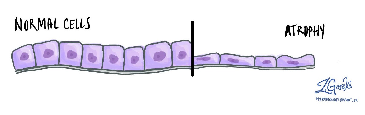
What does atrophic tissue look like under the microscope?
Under the microscope, atrophic tissue looks thinner than normal, with smaller cells and wider spaces between them. These changes indicate that the tissue has adapted to reduced use, limited blood flow, reduced hormonal stimulation, or ongoing inflammation. The symptoms a person notices, if any, depend on which tissue is affected; our article on atrophy describes these in more detail.
Questions to ask your doctor
- What does “atrophic” mean in my pathology report?
- Which tissue or organ is described as atrophic in my case?
- What is causing the atrophic change?
- Is it related to aging, hormone changes, reduced blood flow, or inflammation?
- Does the underlying cause need to be monitored or treated?
- Could this lead to any complications, such as muscle weakness or vitamin deficiencies?
- Will this finding be followed up over time?
Related articles on MyPathologyReport.com
- Atrophy (the noun form, and the main article on this change)
- Hypertrophy (when cells grow larger rather than shrinking)
- Hyperplasia (when the number of cells increases)
- Metaplasia (when one normal cell type is replaced by another)
- Chronic atrophic gastritis (atrophy of the stomach lining)
We are proud to partner with:
![]()