

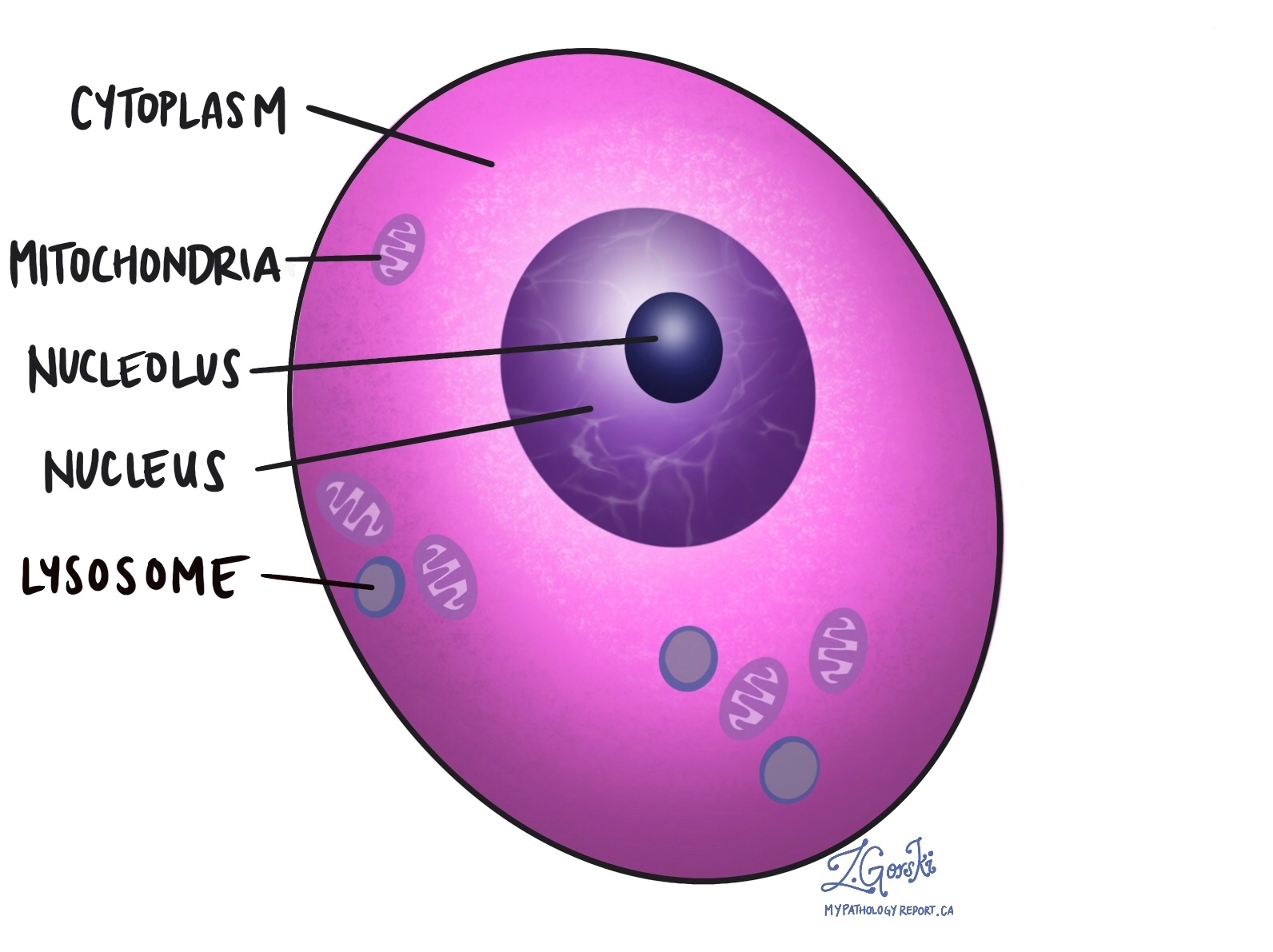
The cytoplasm is the material that fills the inside of a cell, surrounding the nucleus and extending to the cell membrane. It is mostly water and contains proteins, nutrients, and tiny specialized structures called organelles — including mitochondria, which generate energy, and lysosomes, which break down waste. The cytoplasm gives a cell much of its shape and volume, and it is where many of the cell’s essential day-to-day activities take place. In pathology, the appearance of a cell’s cytoplasm under the microscope provides important clues about the cell’s type and whether it appears normal or abnormal.
How do pathologists examine cytoplasm?
Pathologists examine cytoplasm by staining tissue samples with a combination of dyes called hematoxylin and eosin (H&E). Hematoxylin stains the nucleus dark blue or purple, while eosin stains the cytoplasm pink. This color contrast allows the pathologist to clearly see the size, shape, and texture of the cytoplasm in every cell on the slide — and to compare these features between normal cells and potentially abnormal ones.
The amount of cytoplasm also varies considerably between cell types. Squamous cells, which form the flat surface layers of the skin and many internal organs, have abundant cytoplasm that gives them their characteristic broad, flat shape. Lymphocytes — small immune cells found throughout the body — have very little cytoplasm, appearing compact and almost entirely nuclear under the microscope.
How do pathologists describe cytoplasm?
Pathologists use specific terms to describe the color, texture, and content of cytoplasm, which help identify cell types and diagnose conditions. The most commonly used terms include:
- Eosinophilic — the cytoplasm stains bright pink with eosin, indicating a high protein content. This is one of the most common descriptions and is seen in many normal and abnormal cell types.
- Oncocytic — the cytoplasm is densely pink and granular due to an unusually large number of mitochondria packed inside the cell. Oncocytic cells are seen in certain thyroid, salivary gland, and kidney tumors.
- Clear — the cytoplasm appears transparent or pale because it contains fat, glycogen, or other substances that do not take up the standard stains. Clear cytoplasm is a hallmark of certain kidney cancers (clear cell renal cell carcinoma) and some other tumor types.
- Basophilic — the cytoplasm stains blue or purple because it contains substances that attract the hematoxylin stain, often reflecting high RNA content in actively growing or secreting cells.
- Vacuolated — the cytoplasm contains one or more clear spaces called vacuoles, which may represent accumulated fat, mucin, or other material. Vacuolated cytoplasm is seen in some secretory cells and certain tumor types.
- Granular — the cytoplasm contains visible small particles or granules, which may represent secretory products, pigment, or abnormal deposits.
Why does cytoplasm matter in a pathology report?
When your pathology report describes the cytoplasm of cells — for example, “cells with abundant eosinophilic cytoplasm” or “clear cytoplasm” — it is recording features that helped the pathologist identify the cell type and assess whether the cells look normal or abnormal.
Cytoplasm is also relevant in the context of immunohistochemistry (IHC) testing. When a stain is described as showing “cytoplasmic positivity,” it means the marker being tested was detected in the cytoplasm of the cell — an important detail because some markers are normally found in the nucleus, some in the cytoplasm, and some on the cell surface, and an abnormal location can itself be diagnostically significant.
Changes in the amount, color, or texture of the cytoplasm are among the features pathologists use to recognize cancer, identify specific tumor types, and assess how abnormal cells look compared to their normal counterparts.
Questions to ask your doctor
- My report describes a specific type of cytoplasm — what does this tell the pathologist about my diagnosis?
- Is the cytoplasmic appearance of the cells normal for this tissue, or is it an abnormal finding?
Related articles on MyPathologyReport.com
We are proud to partner with:
![]()