by Emily Goebel, MD FRCPC
September 25, 2023
What is adenocarcinoma in situ of the cervix?
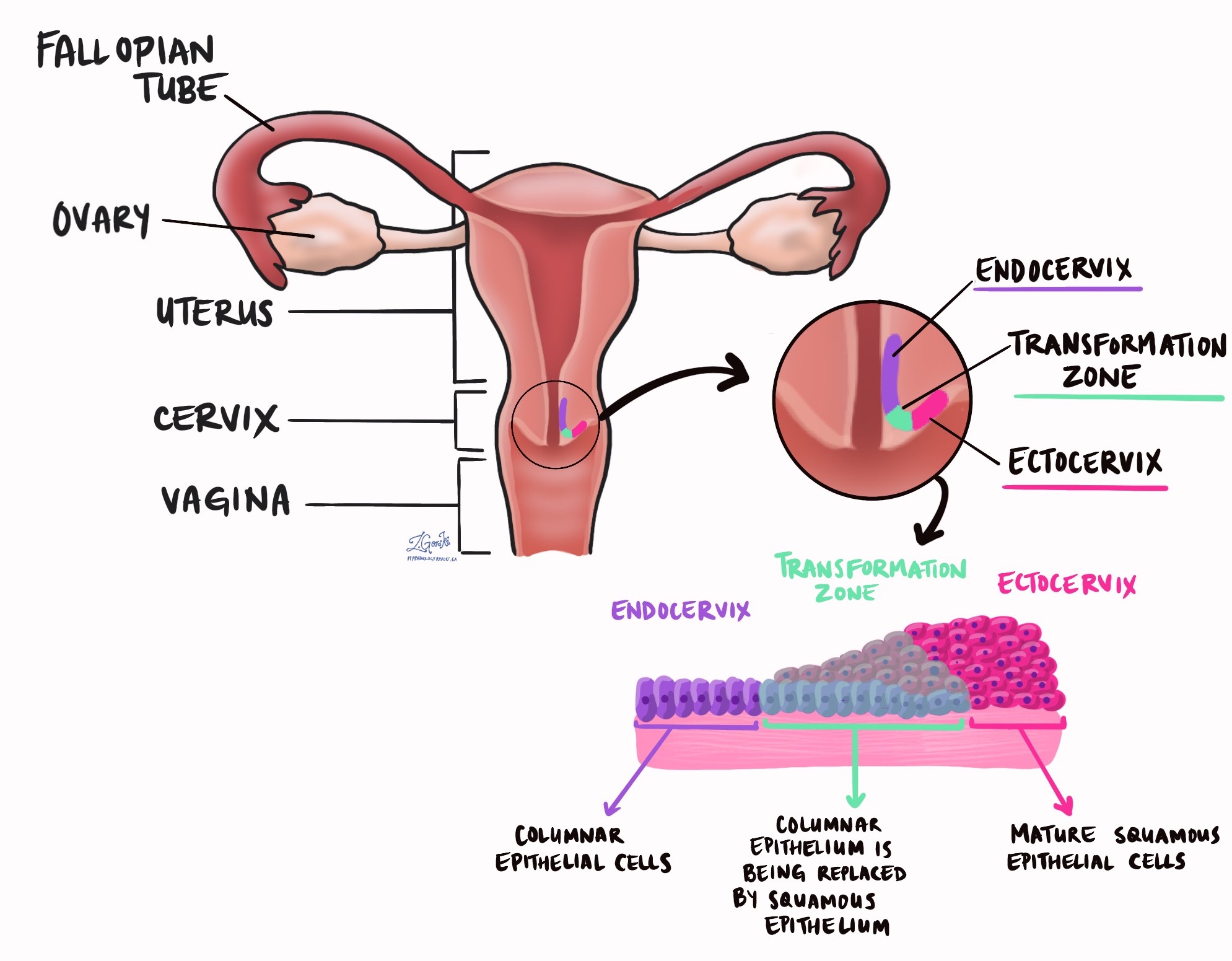
Adenocarcinoma in situ (AIS) is a precancerous disease in the cervix. The disease starts from glands in a part of the cervix called the endocervix. If not treated, AIS can turn into a type of cervical cancer called endocervical adenocarcinoma.

Is adenocarcinoma in situ a type of cervical cancer?
No, AIS is not a type of cervical cancer. However, it is a pre-cancerous condition that can over time turn into a type of cervical cancer called endocervical adenocarcinoma.
What is the difference between adenocarcinoma in situ and endocervical adenocarcinoma?
AIS is a non-invasive condition which means the abnormal cells are only found in a thin layer of tissue called the epithelium on the surface of the cervix. In contrast, in endocervical adenocarcinoma, the tumour cells have spread beyond the epithelium into a layer of tissue called the stroma.
What causes adenocarcinoma in situ in the cervix?
The most common cause of AIS in the cervix is infection by human papillomavirus (HPV). HPV is a sexually transmitted virus that infects cells in the cervix. Once infected, the cells can undergo a series of genetic changes that can lead to the development of AIS. Other conditions caused by HPV in the cervix include endocervical adenocarcinoma, low-grade squamous intraepithelial lesion (LSIL), high-grade squamous intraepithelial lesion (HSIL), and squamous cell carcinoma (SCC).
How do pathologists make the diagnosis of adenocarcinoma in situ in the cervix?
The diagnosis of AIS is usually made after some cells are removed from the cervix during a procedure called a Pap test. The diagnosis can also be made after a larger sample of tissue is removed in a biopsy or excision. The tissue is then examined under a microscope by a pathologist.

What is p16 and why is it important?
Cells infected with high-risk types of human papillomavirus (HPV) produce large amounts of a protein called p16. Your pathologist may perform a test called immunohistochemistry to look for p16 inside the abnormal cells. This test will confirm the diagnosis of AIS and rule out other conditions that can look like AIS under the microscope. Almost all cases of AIS are positive or reactive for p16 which means your pathologist saw the p16 protein in the cancer cells.
What is a margin?
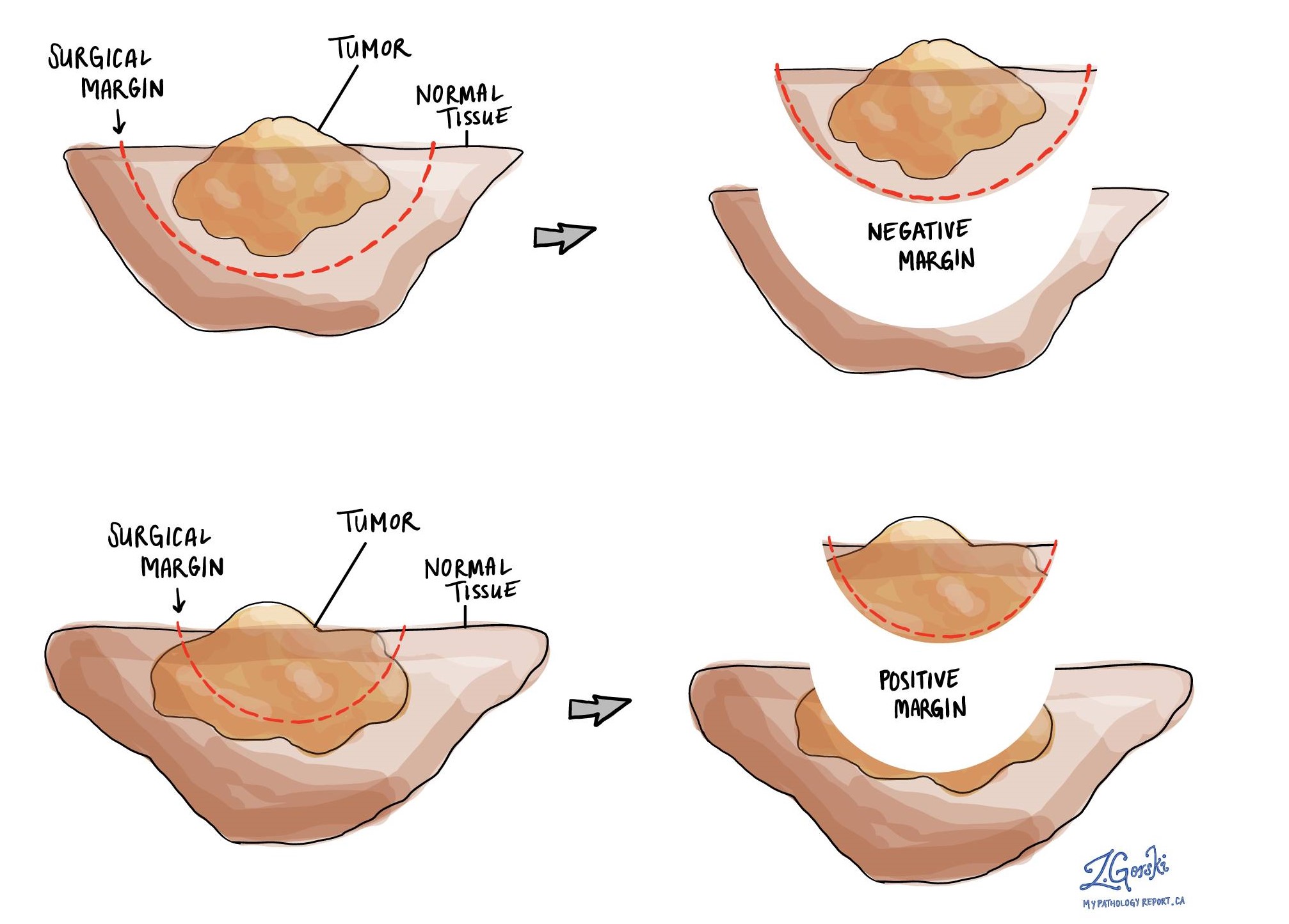
A margin is any tissue that has to be cut by the surgeon in order to remove the tumour from your body. If you underwent a surgical procedure to remove the entire tumour from your body, your pathologist will examine the margin closely to make sure there are no cancer cells at the cut edge of the tissue. A margin is considered positive when AIS is seen at the edge of the cut tissue. Finding AIS at the margin increases the risk that the tumour will grow back in that location.

The number and type of margins described in your report will depend on the type of procedure performed to remove the tumour from your body. Pap smears and small biopsy tissue samples do not have margins
Typical margins include:
- Endocervical margin – This is where the cervix meets the inside of the uterus.
- Ectocervical margin – This is the bottom of the cervix, closest to the vagina.
- Stromal margin – This is the tissue inside the wall of the cervix.


