by Jason Wasserman MD PhD FRCPC
July 22, 2024
Background:
Angioleiomyoma is a noncancerous tumour made up of abnormal blood vessels. While it can be found anywhere in the body, it most commonly involves the skin or the deep soft tissue below it. Another name for this tumour is vascular leiomyoma.
What are the symptoms of angioleiomyoma?
The most common symptom of an angioleiomyoma is pain when pressure is applied to the tumour.
What causes an angioleiomyoma?
What causes angioleiomyoma is currently unknown. Some evidence suggests, however, that prior trauma can cause this tumour.
How is the diagnosis made?
The diagnosis can be made after the tumour is removed and the tissue is examined under a microscope by a pathologist.
Microscopic features of this tumour
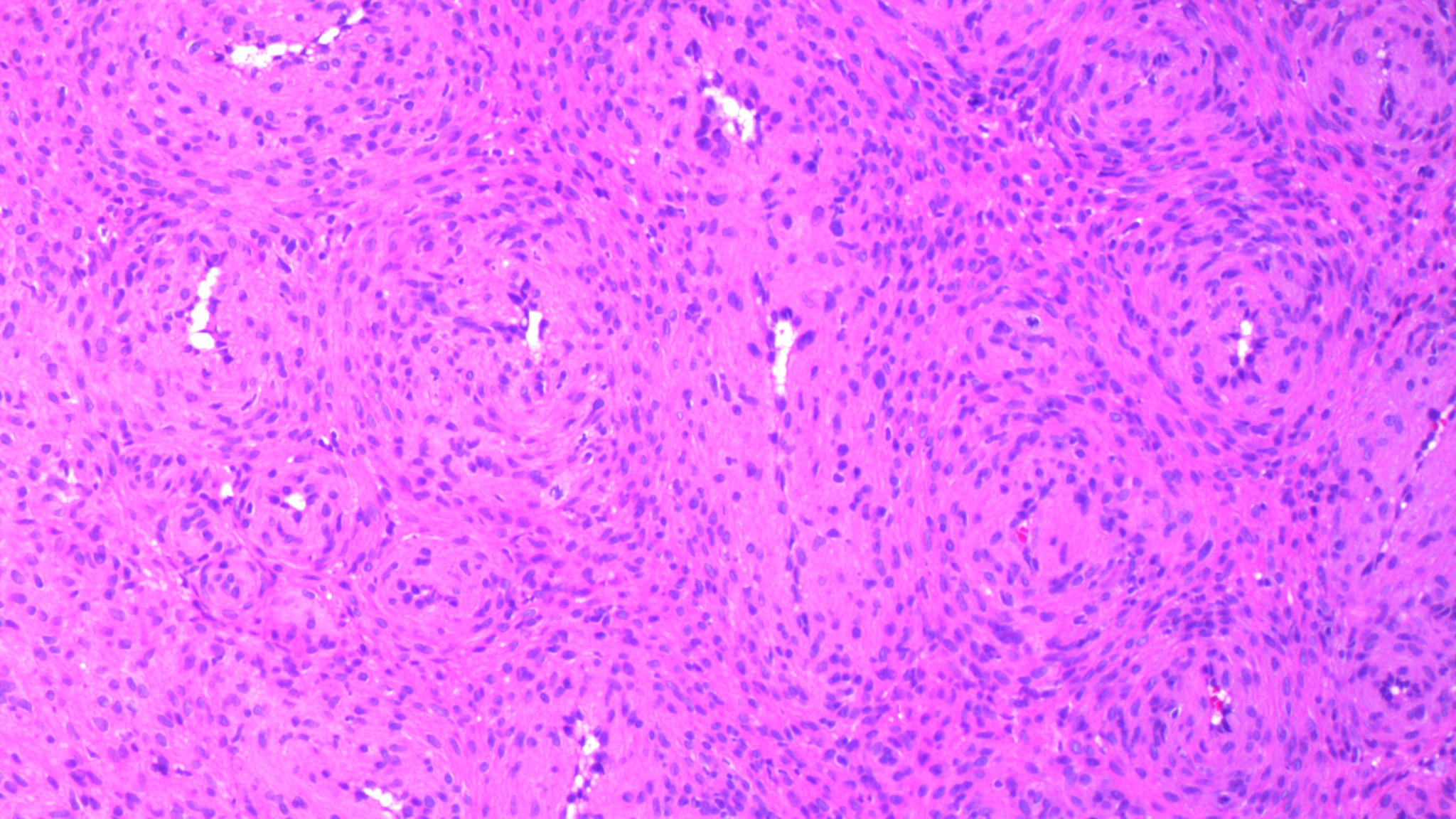
When examined under the microscope, the tumour is made up of variably sized blood vessels surrounded by long, thin spindle-shaped cells. Depending on the shape and size of the blood vessels in the tumour, some pathologists subtype the tumour as solid, venous, and cavernous. All subtypes are noncancerous and the subtype does not change the behavior of the tumour over time.

Immunohistochemistry
Pathologists often order a test called immunohistochemistry (IHC) to confirm the diagnosis and to rule out other conditions that can look like angioleiomyoma under the microscope. When IHC is performed, the tumour is typically positive or reactive for smooth muscle markers including smooth muscle actin, caldesmon and calponin. Some tumours are also positive for the muscle marker desmin.
About this article
Doctors wrote this article to help you read and understand your pathology report. If you have additional questions, contact us.


