Jason Wasserman MD PhD FRCPC
July 3, 2025
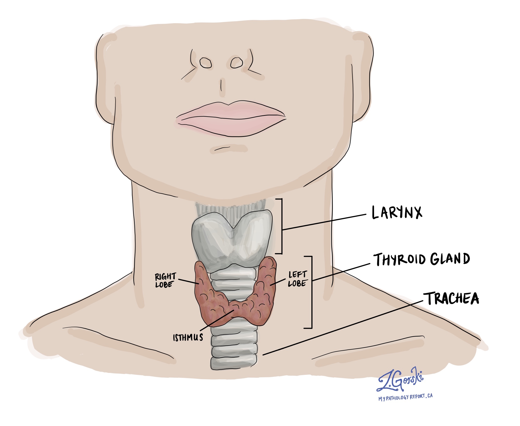
A benign follicular nodule is a non-cancerous lump that forms in the thyroid gland. The thyroid is a small, butterfly-shaped gland located in the front of the neck. It produces hormones that help regulate your metabolism, energy levels, and many important body functions.

The word “benign” means that the nodule is not cancerous and does not have the potential to spread to other parts of the body. Benign follicular nodules are common and often develop as part of a condition known as nodular thyroid hyperplasia. In this condition, the thyroid becomes enlarged and contains multiple nodules of different sizes.
What causes a benign follicular nodule?
In many cases, the exact cause of a benign follicular nodule is not known. However, several factors may increase the chance of developing thyroid nodules:
-
Iodine deficiency: Iodine is an important mineral that the thyroid uses to make hormones. A lack of iodine in the diet can lead to thyroid enlargement and the formation of nodules.
-
Genetic factors: Some individuals may be more susceptible to developing nodules due to inherited traits or a family history of the condition.
-
Hormonal influences: Changes in hormone levels, such as those that occur during pregnancy or menopause, may also play a role.
-
Chronic inflammation: Conditions like Hashimoto’s thyroiditis, an autoimmune disease, may cause the thyroid to become inflamed and develop nodules.
Despite these possible causes, many benign nodules develop without a clear reason.
How is this diagnosis made?
The process of diagnosing a benign follicular nodule typically involves several steps:
-
Physical examination: A doctor may feel a lump in the neck during a routine check-up or after a patient reports symptoms.
-
Imaging tests: An ultrasound is commonly used to get a detailed image of the thyroid and assess the size, shape, and appearance of the nodule.
-
Fine-needle aspiration biopsy (FNAB): This is the most important test for determining whether a thyroid nodule is benign or suspicious. A thin needle is used to collect a small sample of cells from the nodule, usually under ultrasound guidance. The sample is then examined under a microscope by a pathologist.
What does a benign follicular nodule look like under the microscope?
When viewed under a microscope, a benign follicular nodule is composed of follicular cells, the same type of cells that are normally found in the thyroid gland. These cells are typically arranged in small, round groups called follicles, which often contain a thick, pink fluid called colloid.
The follicular cells in a benign nodule are usually small, evenly spaced, and show no signs of abnormal growth. There is little to no crowding or overlapping of the cells, which helps pathologists distinguish a benign nodule from cancer.
In some cases, inflammatory cells such as lymphocytes (a type of white blood cell) or histiocytes (a type of immune cell) may also be present, especially if the nodule is associated with chronic lymphocytic thyroiditis.
What happens after a fine-needle aspiration biopsy?
If the biopsy results show a benign follicular nodule, no immediate treatment is usually necessary. Most benign nodules are monitored over time with regular check-ups and follow-up imaging to make sure they are not growing or changing in appearance.
Surgery may be recommended in certain situations, such as:
-
The nodule is causing pressure, pain, or discomfort.
-
It affects swallowing or breathing.
-
It continues to grow or changes in appearance.
-
There is uncertainty in the biopsy result or concern for cancer based on other clinical findings.
Your doctor will help decide the best approach based on your specific situation, symptoms, and preferences.
What is the risk of cancer?
The risk of cancer in a benign follicular nodule is very low. A diagnosis of “benign” after a fine-needle aspiration biopsy is considered reliable, and most people do not require surgery or further invasive testing. However, because no test is perfect, doctors may recommend regular monitoring to watch for any changes. This may include repeat ultrasounds and, in some cases, a second biopsy if the nodule grows or develops new features.
Questions to ask your doctor
- How often should I have follow-up imaging?
-
Is there any chance this nodule could turn into cancer?
-
Would surgery be necessary in my case?
-
Are there symptoms I should watch for?
-
Is this nodule affecting my thyroid function?


