by Jason Wasserman MD PhD FRCPC
August 22, 2023
What is a glomus tumour?
A glomus tumour is an abnormal growth made up of cells from the glomus body, a structure normally found on the outside of small blood vessels.
Is a glomus tumour benign or malignant?
The vast majority of glomus tumours are benign (noncancerous). Although exceedingly rare, malignant (cancerous) glomus tumours can occur.
Can a glomus tumour become cancerous over time?
In very rare cases, a previously benign (noncancerous) glomus tumour can become malignant (cancerous) over time. Unlike the cells in a benign glomus tumour, the cells in a malignant glomus tumour can metastasize (spread) to other parts of the body.
What causes a glomus tumour?
Most glomus tumours are sporadic which means they are not associated with any known genetic syndrome. More than half of all sporadic glomus tumours will contain a genetic alteration involving one of the genes in the NOTCH family. At this time, doctors do not know what causes this genetic alteration to occur. Genetic syndromes associated with the development of multiple glomus tumours include multiple familial glomus tumours syndrome and neurofibromatosis type-1.
What are the symptoms of a glomus tumour?
The symptoms of a glomus tumour depend on the size and location of the tumour. Glomus tumours in the skin are painful when touched or exposed to the cold. Deep soft tissue tumours or those within an internal organ may not cause any symptoms until the tumour becomes large enough to put pressure on surrounding tissues.
Where are glomus tumours found in the body?
Glomus tumours can develop anywhere in the body; however, most are found in the skin on the hands and feet. Other common locations include deep soft tissue, the digestive tract (especially the stomach), and the genitourinary tract.
How is this diagnosis made?
The diagnosis of glomus tumour after the tumour is removed and sent to a pathologist for examination under the microscope.
What does a glomus tumour look like under the microscope?
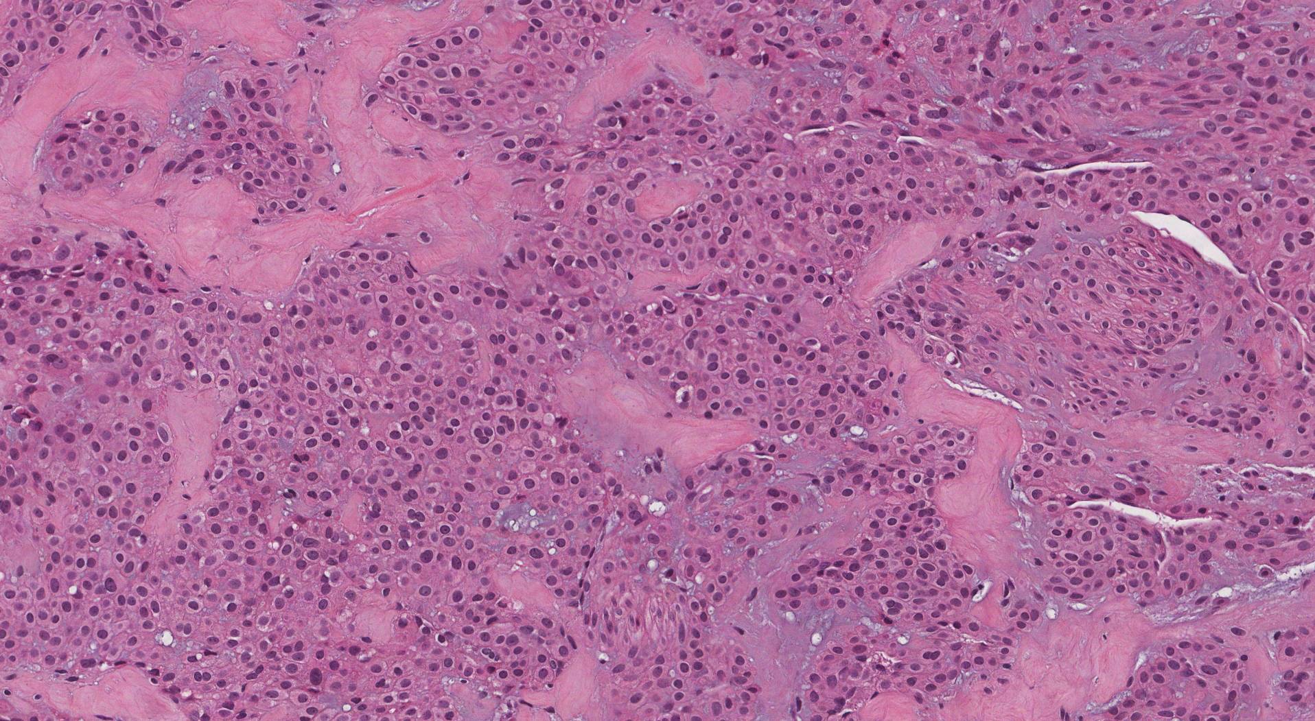
When examined under the microscope, a glomus tumour is made up of small round light pink cells with centrally placed nuclei and well-defined cell borders. The cells may be described as uniform or monotonous because they all look very similar to one another. The tumour cells are typically arranged in large groups called nests that surround small blood vessels. Mitotic figures (cells dividing to create new cells) are rare.

What often tests may be performed to confirm the diagnosis?
Pathologists often perform a test called immunohistochemistry (IHC) to confirm the diagnosis of a glomus tumour. When this test is performed, the tumour cells are typically positive for markers expressed by normal glomus cells including smooth muscle actin (SMA), h-caldesmon, and collagen IV.


