by Jason Wasserman MD PhD FRCPC
April 28, 2023
What is squamous cell carcinoma of the oropharynx?
Squamous cell carcinoma (SCC) is the most common type of oropharyngeal cancer. The oropharynx is an area of the throat that includes the tonsils, base of the tongue, uvula, and soft palate. This type of cancer quickly spreads to lymph nodes especially those in the neck. For many patients, the first sign of the disease is a noticeable lump in the neck.

What causes squamous cell carcinoma in the oropharynx?
Most SCCs in the oropharynx are caused by long-standing infection with human papillomavirus (HPV). Less common causes of SCC in the oropharynx are smoking, excessive alcohol consumption, and immune suppression.
How is this diagnosis made?
The diagnosis of SCC in the oropharynx is usually made after a small tissue sample is removed in a procedure called a biopsy. The biopsy may be taken from the oropharynx or it may be taken from the neck. For some patients, surgery may be performed to remove the entire tumour. Other patients may receive radiation therapy with or without surgery to remove the tumour.
If the tumour is removed, it will be sent to a pathologist who will prepare another pathology report. This report will confirm or revise the original diagnosis and provide additional important information such as tumour size and the spread of tumour cells to lymph nodes. This information is used to determine the cancer stage and to decide if additional treatment is required.
What does it mean if squamous cell carcinoma of the oropharynx is described as nonkeratinizing?
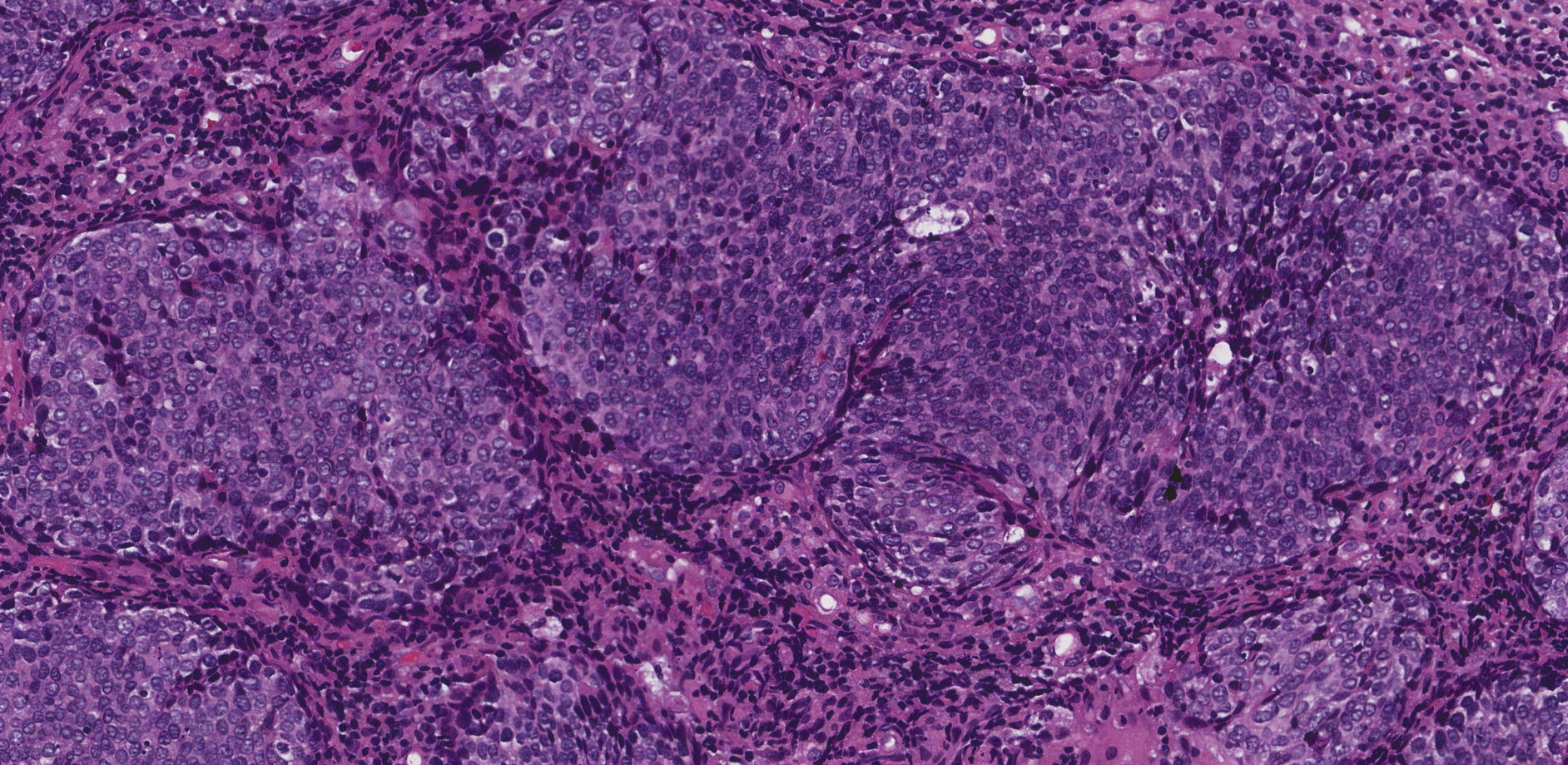
Nonkeratinizing is a term pathologists use to describe cells that look purple/blue when examined under the microscope because the cytoplasm (body) of the cell contains very little of a specialized protein called keratin. These types of cells are sometimes described as basaloid because they look similar to normal basal cells. Most SCCs of the oropharynx caused by HPV are nonkeratinizing.

What does it mean if squamous cell carcinoma of the oropharynx is described as keratinizing?
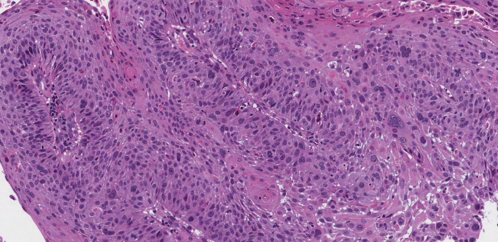
Keratinizing is a term pathologists use to describe cells that look pink when examined under the microscope because the cytoplasm (body) of the cell contains large amounts of a specialized protein called keratin. Some SCCs of the oropharynx, in particular those not caused by HPV, are keratinizing.

What is p16 and why is it important?
Squamous cells infected with HPV produce a large amount of a protein called p16 which pathologists can see using a test called immunohistochemistry. Tumours made up of cells that produce extra p16 are described as positive or reactive while those that do not produce extra p16 are reported as negative or non-reactive. Most tumours in the oropharynx will be positive for p16.
What does metastatic squamous cell carcinoma mean?
Metastatic SCC means that cancer cells have spread from the tumour to another organ or tissue such as a lymph node. It is very common for SCC of the oropharynx to spread to lymph nodes in the neck and for many patients, the first sign of disease will be a lump on the front or side of the neck.
How is the size of the tumour measured and why is it important?
The tumour size can only be determined after the entire tumour has been removed surgically from the throat. The tumour size is important because it is used to determine the tumour stage (pT) and because larger tumours are associated with a worse prognosis.
What is a margin and why are margins important?
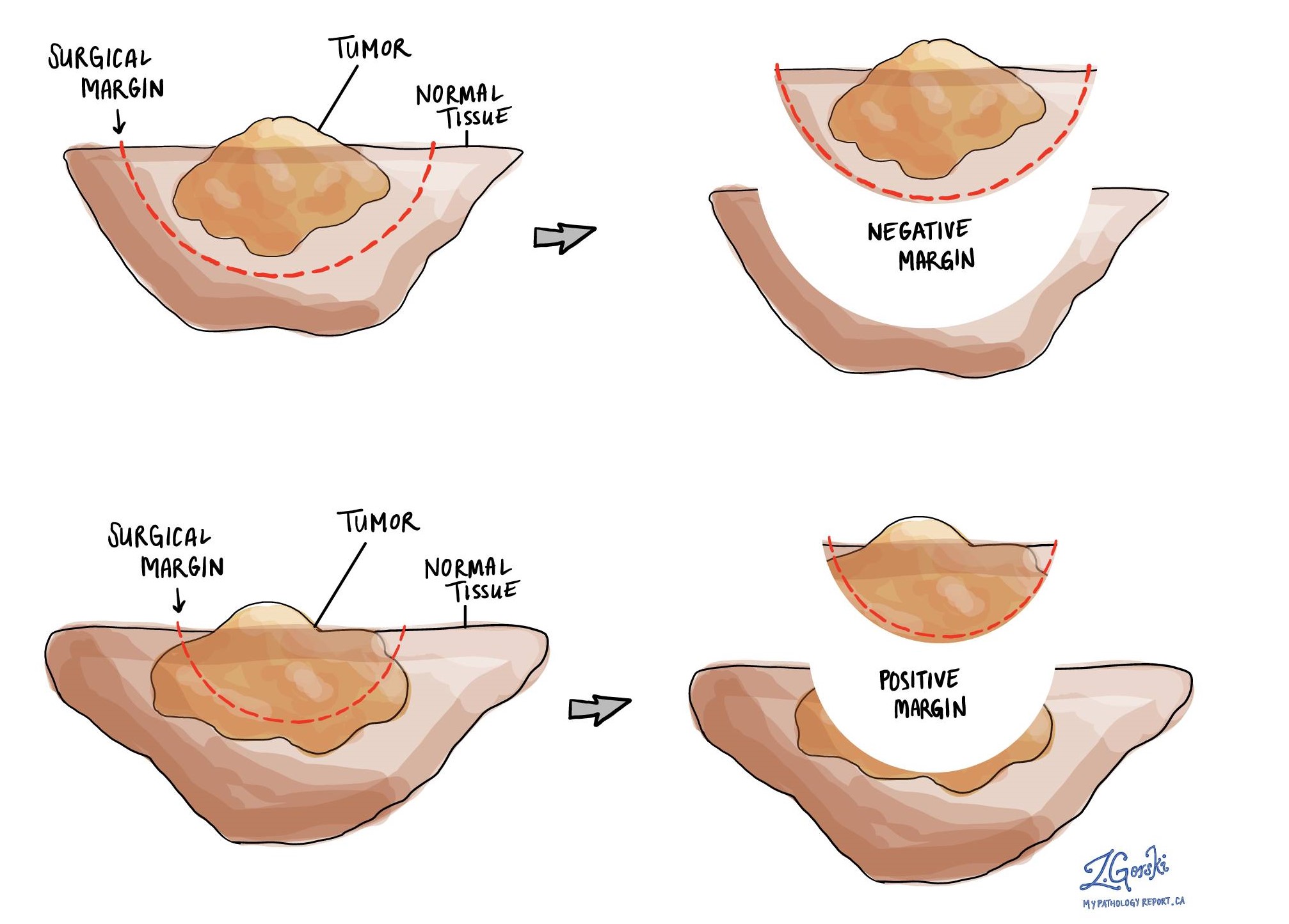
A margin is any tissue that was cut by the surgeon in order to remove the tumour from your body. The types of margins described in your report will depend on the organ involved and the type of surgery performed. Margins will only be described in your report after the entire tumour has been removed in an excision or resection. Margins are typically not described for biopsy specimens.
A negative margin means that no tumour cells were seen at any of the cut edges of the tissue. A margin is called positive when there are tumour cells at the very edge of the cut tissue. A positive margin is associated with a higher risk that the tumour will recur in the same site after treatment.


