by Jason Wasserman MD PhD FRCPC
October 7, 2022
What is high-grade serous carcinoma of the ovary?
High-grade serous carcinoma is the most common type of ovarian and fallopian tube cancer. It starts from cells normally found on the outside surface of the ovary or the inside surface of the fallopian tube. Regardless of where the tumour starts, it often involves both organs at the time of diagnosis. It is common for high-grade serous carcinoma to quickly spread to other organs inside the abdomen and pelvis and to the peritoneum, a thin layer of tissue that covers these organs.
What causes high-grade serous carcinoma of the ovary?
At present doctors do not know what causes high-grade serous carcinoma. However, risk factors associated with this type of cancer include a family or personal history of breast or ovarian cancer and infertility.
How is this diagnosis made?
The diagnosis of high-grade serous carcinoma can also be made after a small sample of tissue is removed in a procedure called a biopsy. In this procedure, a small sample of tissue from the pelvis or abdomen is removed. The ovary itself is not usually biopsied.
Because it often spreads to the peritoneum, high-grade serous carcinoma can also be diagnosed after fluid is removed from the abdominal cavity in a procedure called a fine needle aspiration (FNA). The fluid is then sent to a pathologist who examines the cells in the fluid under the microscope.
For some women, the diagnosis of high-grade serous carcinoma is only made when the entire tumour has been surgically removed and sent to a pathologist for examination. The ovary is usually removed along with the fallopian tube, and uterus.
Your surgeon may request an intraoperative or frozen section consultation from your pathologist. The diagnosis made by your pathologist during the intraoperative consultation can change the type of surgery performed or the treatment offered after the surgery is completed.
Why is it important if the tumour was received intact or ruptured?
All ovarian tumours are examined to see if there are any holes or tears in the outer surface of the tumour or ovary. The outer surface is referred to as the capsule. The capsule is described as intact if no holes or tears are identified. The capsule is described as ruptured if the outer surface contains any large holes or tears.
This information is important because a capsule that ruptures inside the body may spill cancer cells into the abdominal cavity. A ruptured capsule is associated with a worse prognosis and is used to determine the tumour stage.
Were cancer cells seen on the surface of the ovary or fallopian tube?
The cancer cells in high-grade serous carcinoma can spread from the ovary to another nearby organ such as the fallopian tube or the ovary on the other side of the body. If cancer cells are seen on the surface of the fallopian tube or ovary, it suggests that they have travelled there from another site. This information is important because a tumour that has spread from one organ to another is given a higher tumour stage.
What are peritoneal implants and why are they important?
Peritoneal implants are groups of cancer cells that have spread to the peritoneum, a thin layer of tissue that covers the organs inside of the pelvis and abdomen. Tissue from the peritoneum is often sent along with the ovaries, fallopian tubes, and uterus so they can be examined by your pathologist for cancer cells.
Unfortunately, it is common for high-grade serous carcinoma to spread to the peritoneum. Peritoneal implants are associated with a worse prognosis and are used to determine the tumour stage.
Has the tumour spread to other organs or tissues in the pelvis or abdomen?
Small samples of tissue are commonly removed in a procedure called a biopsy to see if tumour cells have spread outside of the ovary. These biopsies, which are often from a tissue in the pelvis and abdomen called the peritoneum, are sent to your pathologist to see if the tumour has spread or metastasized. The omentum is an abdominal organ that is a common site of tumour spread or metastasis. This organ is often entirely removed and examined by your pathologist. Other organs (such as the bladder, small intestine, or large intestine) are not typically removed and sent for pathological examination unless they are directly attached to the tumour or tumour spread to these organs is seen by your surgeon. In these cases, your pathologist will examine each organ under the microscope to see if there are any cancer cells attached to those organs. The presence of tumour cells in other organs is used to determine the tumour (T) stage and distant metastatic disease (M) stage.
Were lymph nodes examined and did any contain cancer cells?
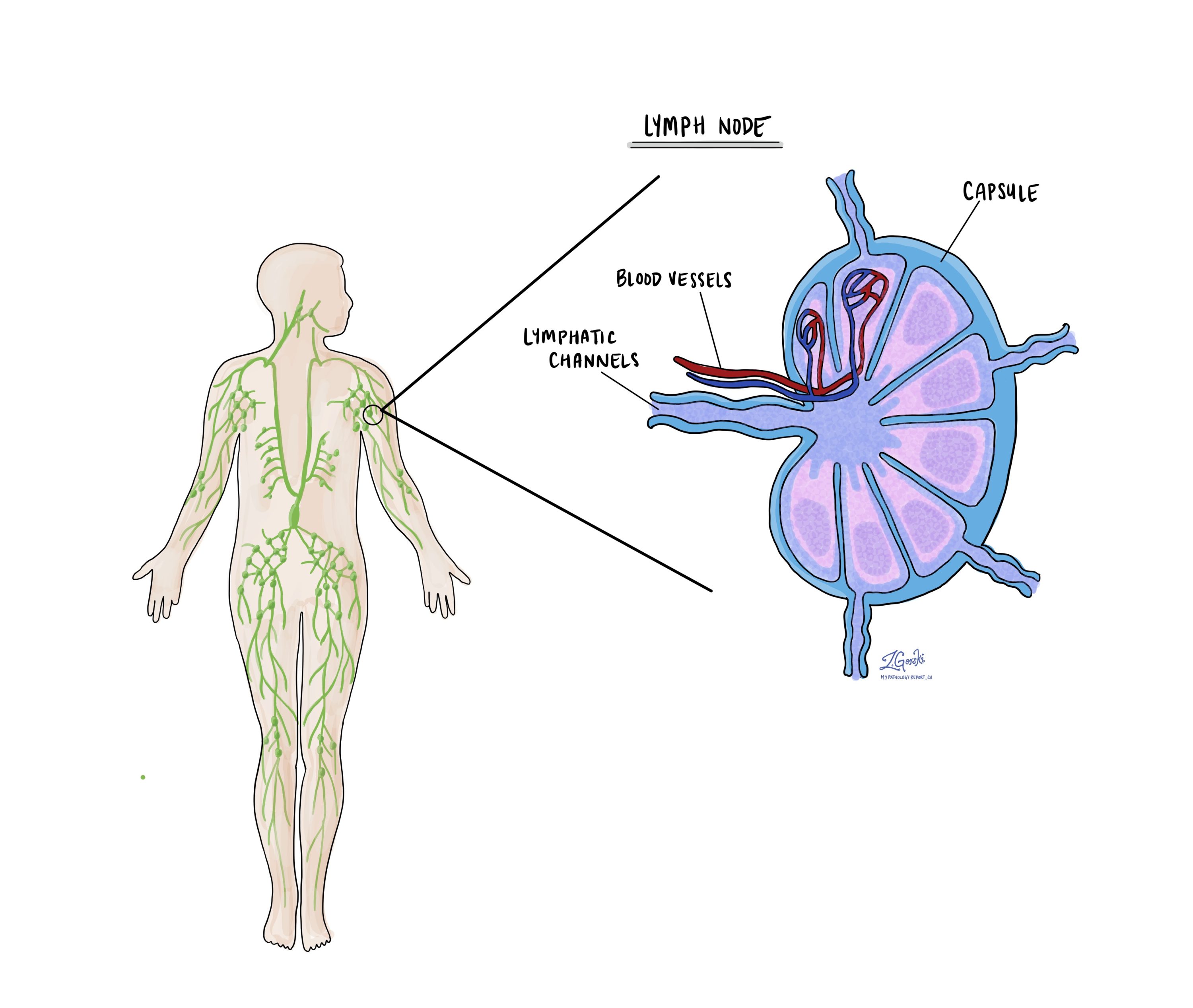
Lymph nodes are small immune organs found throughout the body. Cancer cells can spread from a tumour to lymph nodes through small vessels called lymphatics. For this reason, lymph nodes are commonly removed and examined under a microscope to look for cancer cells. The movement of cancer cells from the tumour to another part of the body such as a lymph node is called a metastasis.
Cancer cells typically spread first to lymph nodes close to the tumour although lymph nodes far away from the tumour can also be involved. For this reason, the first lymph nodes removed are usually close to the tumour. Lymph nodes further away from the tumour are only typically removed if they are enlarged and there is a high clinical suspicion that there may be cancer cells in the lymph node.
If any lymph nodes were removed from your body, they will be examined under the microscope by a pathologist and the results of this examination will be described in your report. Most reports will include the total number of lymph nodes examined, where in the body the lymph nodes were found, and the number (if any) that contain cancer cells. If cancer cells were seen in a lymph node, the size of the largest group of cancer cells (often described as “focus” or “deposit”) will also be included.
The examination of lymph nodes is important for two reasons. First, this information is used to determine the pathologic nodal stage (pN). Second, finding cancer cells in a lymph node increases the risk that cancer cells will be found in other parts of the body in the future. As a result, your doctor will use this information when deciding if additional treatment such as chemotherapy, radiation therapy, or immunotherapy is required.

What does it mean if a lymph node is described as positive?
Pathologists often use the term “positive” to describe a lymph node that contains cancer cells. For example, a lymph node that contains cancer cells may be called “positive for malignancy” or “positive for metastatic carcinoma”.
What does it mean if a lymph node is described as negative?
Pathologists often use the term “negative” to describe a lymph node that does not contain any cancer cells. For example, a lymph node that does not contain cancer cells may be called “negative for malignancy” or “negative for metastatic carcinoma”.
What are isolated tumour cells (ITCs)?
Pathologists use the term ‘isolated tumour cells’ to describe a group of tumour cells that measures 0.2 mm or less and is found in a lymph node. Lymph nodes with only isolated tumour cells (ITCs) are not counted as being ‘positive’ for the purpose of the pathologic nodal stage (pN).
What is a micrometastasis?
A ‘micrometastasis’ is a group of tumour cells that measures from 0.2 mm to 2 mm and is found in a lymph node. If only micrometastases are found in all the lymph nodes examined, the pathologic nodal stage is pN1mi.
What is a macrometastasis?
A ‘macrometastasis’ is a group of tumour cells that measures more than 2 mm and is found in a lymph node. Macrometastases are associated with a worse prognosis and may require additional treatment.
What does extranodal extension mean?
All lymph nodes are surrounded by a thin layer of tissue called a capsule. Extranodal extension means that cancer cells within the lymph node have broken through the capsule and have spread into the tissue outside of the lymph node. Extranodal extension is important because it increases the risk that the tumour will regrow in the same location after surgery. For some types of cancer, extranodal extension is also a reason to consider additional treatment such as chemotherapy or radiation therapy.
What does treatment effect mean?
If you were treated with chemotherapy (or other drugs designed to kill cancer cells) prior to surgical removal of the tumour, your pathologist will examine the tumour to determine the percentage of the tumour that is still viable (living tumour cells).
The response will be categorized as follows:
- No/minimal response – Most of the tumour is viable.
- Appreciable response – Some of the tumour is dead and some is viable.
- Complete response – Almost all or all of the tumour is dead.


