

by Jason Wasserman MD PhD FRCPC and Aurelia Busca MD PhD FRCPC
May 15, 2023
What is a serous borderline tumour?
Serous borderline tumour is a non-cancerous type of ovarian tumour. Although it is considered non-cancerous, the tumour cells can spread to other organs within the abdomen and pelvis.

Is a serous borderline tumour of the ovary a type of cancer?
No. Serous borderline tumour is not a type of cancer. However, this tumour has the potential to act in a more aggressive manner than a typical non-cancerous tumour. For example, the tumour cells can spread to other organs in the pelvis and abdomen. For that reason, it is described as “borderline”.
What are the symptoms of a serous borderline tumour?
Symptoms of a serous borderline tumour include abdominal or pelvic pain, swelling, distension, and bloating.
What causes a serous borderline tumour?
At present, the cause of serous borderline tumour remains unknown.
How is the diagnosis of serous borderline tumour made?
For most women, the diagnosis of serous borderline tumour is only made when the entire tumour has been surgically removed and sent to a pathologist for examination. The fallopian tube and uterus may be removed at the same time.
Your surgeon may request an intraoperative or frozen section consultation from your pathologist. The diagnosis made by your pathologist during the intraoperative consultation can change the type of surgery performed or the treatment offered after the surgery is completed.
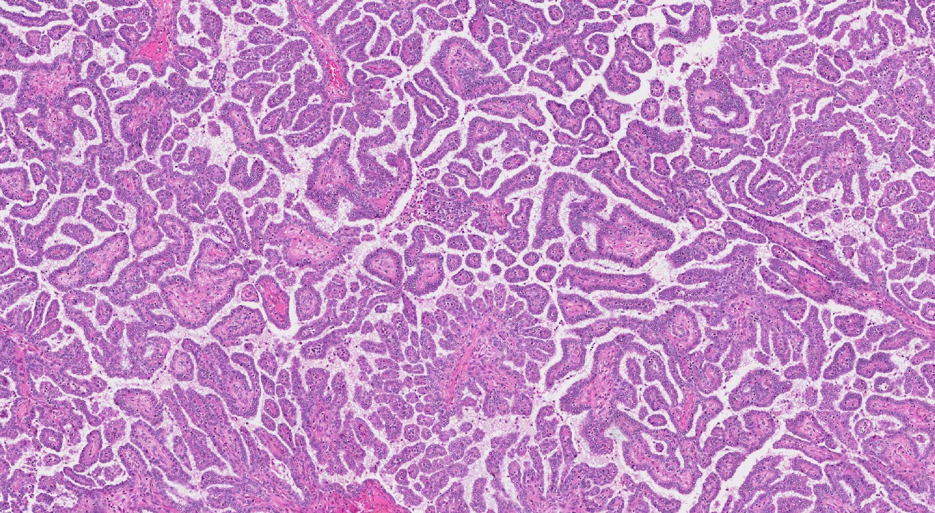
What does a serous borderline tumour look like under the microscope?
Serous borderline tumours develop from the epithelial cells on the outer surface of the ovary. The tumour is usually made up of many small spaces. Pathologists call these spaces cysts. The walls of the cysts can be thin or thick and more solid areas may be found inside some of the cysts. Pathologists sometimes call these solid areas excrescences.
The inside of the cysts are lined by specialized serous cells. These cells produce a clear fluid that fills the inside of the tumour. The cysts with thick walls and the solid areas may have small finger-like projections of tissue. Pathologists call these projections papillary. The more solid areas and papillary projections are what make a serous borderline tumour different from a serous cystadenoma.
What does it mean if the tumour is described as being intact or ruptured?
All ovarian tumours are examined to see if there are any holes or tears in the outer surface of the tumour or ovary. The outer surface is referred to as the capsule. The capsule is described as intact if no holes or tears are identified. The capsule is described as ruptured if the outer surface contains any large holes or tears.
What are non-invasive implants are why are they important?
Pathologists use the term non-invasive implants to describe groups of tumour cells from a serous borderline tumour that have attached to an organ within the abdomen or pelvis. Organs commonly involved by non-invasive implants include the omentum and peritoneum. Unlike “invasive” implants, “non-invasive” implants are only found on the outside surface of an organ. Non-invasive implants are important because they increase the pathologic tumour stage and are associated with an increased risk of tumour regrowth in the abdomen or pelvis.
What does it mean if a serous borderline tumour is described as microinvasive or showing microinvasion?
The tumour cells in a serous borderline tumour are typically only found in the thin layer of tissue that connects with the outside surface of the ovary. However, in a small percentage of tumours, the tumour cells spread into the tissue below the surface. This tissue is called the stroma and the spread of tumour cells into the stroma is called “microinvasion”. In order to be defined as microinvasion, the group of tumour cells within the stroma must be smaller than 5 mm. If the area of microinvasion is equal to or greater than 5 mm, the tumour should be diagnosed as low grade serous carcinoma.
Were lymph nodes examined and did any contain tumour cells?
Lymph nodes are small immune organs located throughout the body. Although rare, the tumour cells from a serous borderline tumour can spread to lymph nodes. If tumour cells are found in a lymph node it will be described in your pathology report. Unlike other types of tumours, cells from a serous borderline tumour found in a lymph node are not associated with a worse prognosis.

We are proud to partner with:
![]()